44 eye anatomy
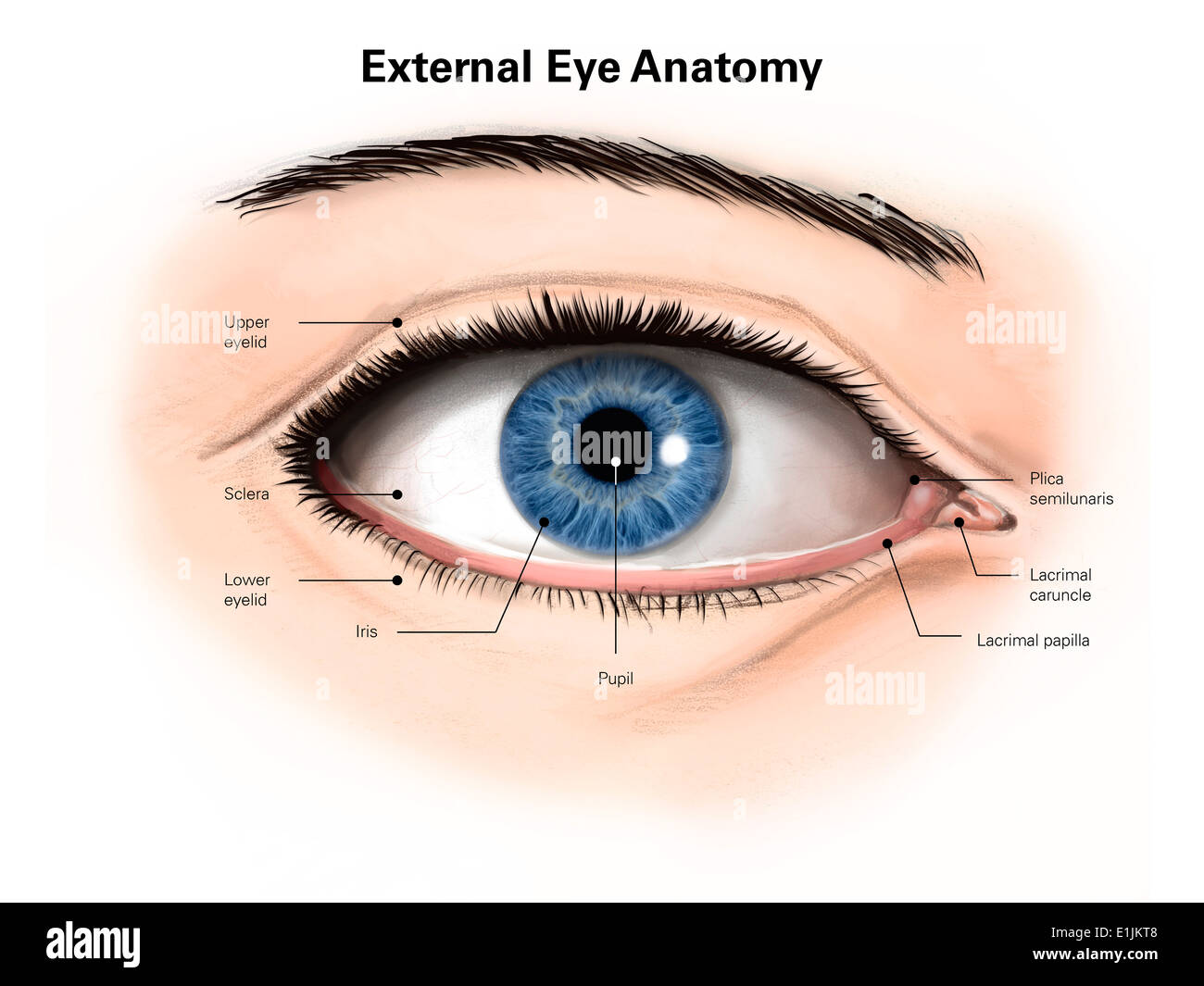
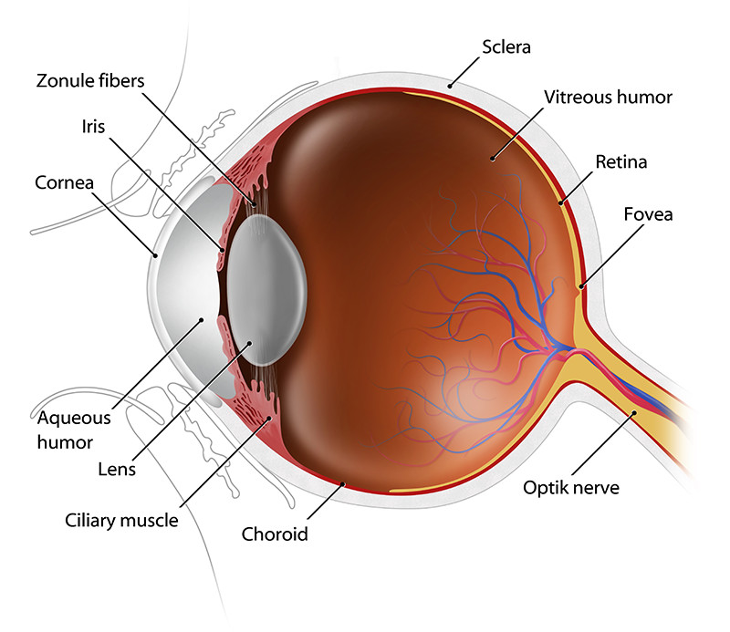
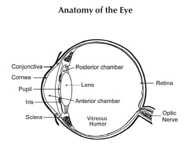
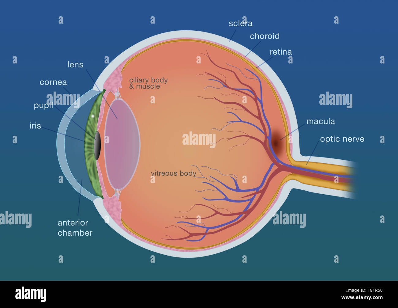
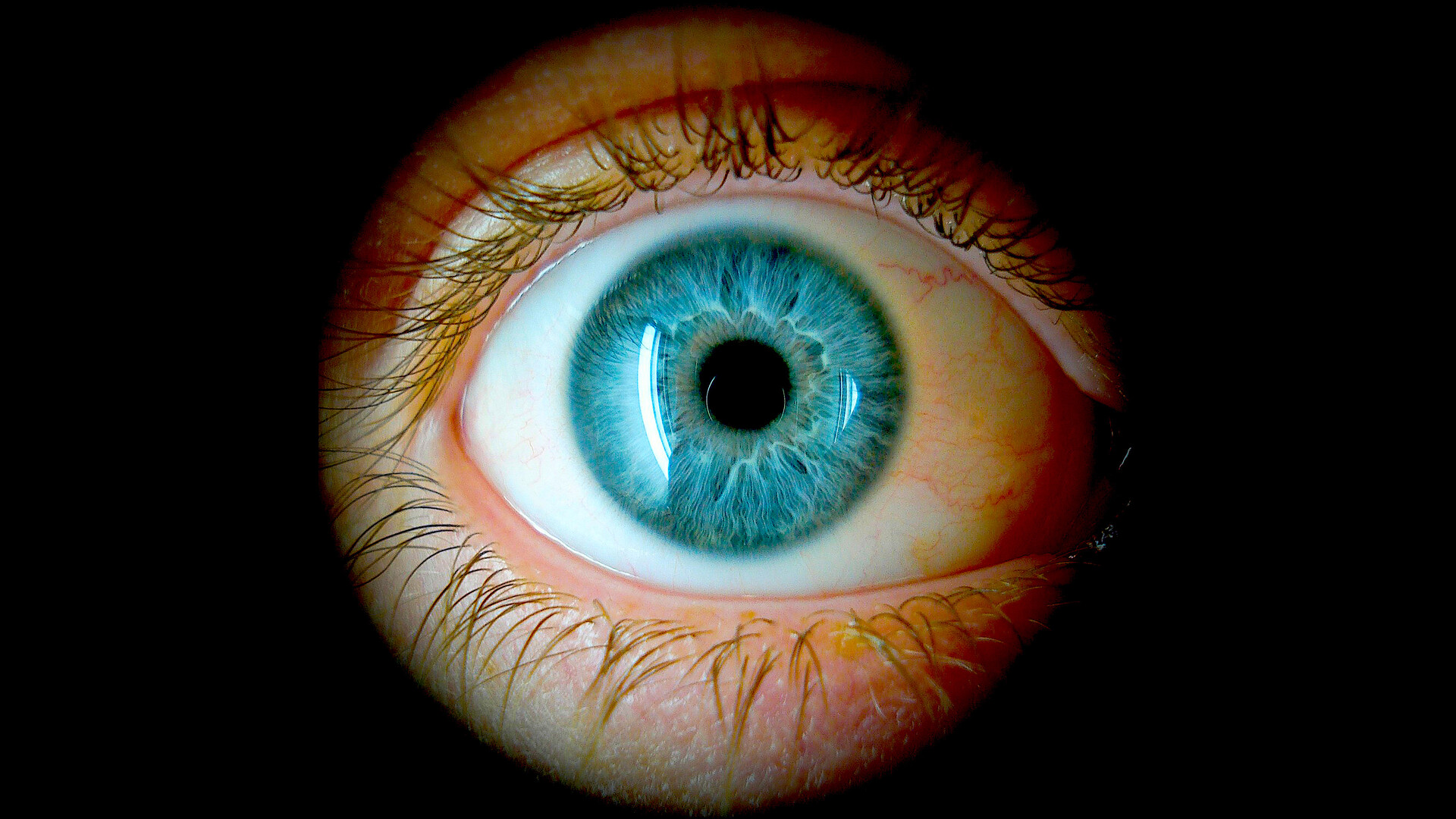
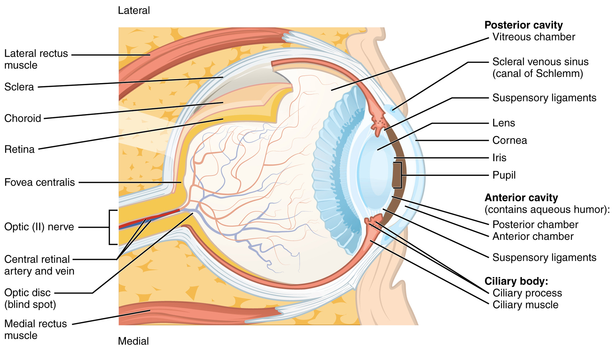
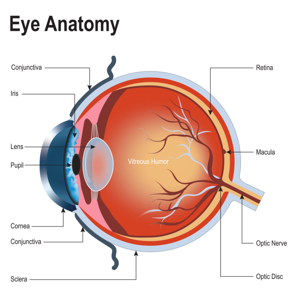
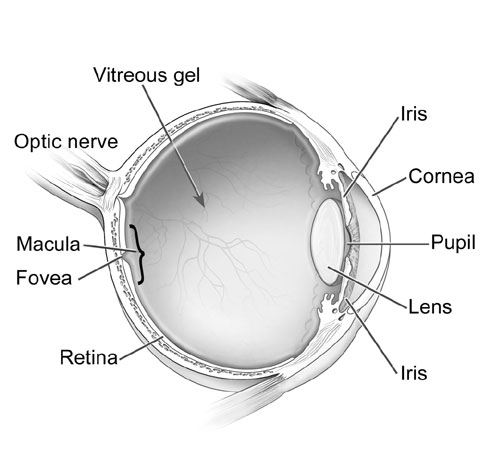
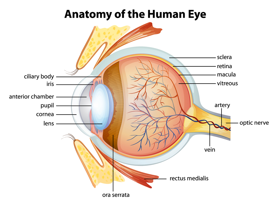
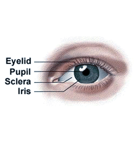
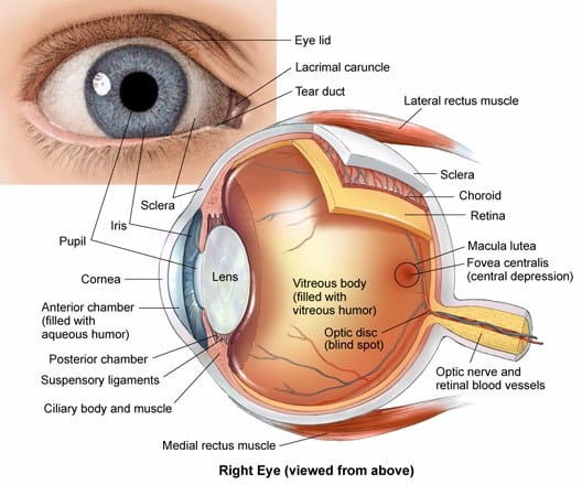
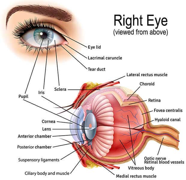
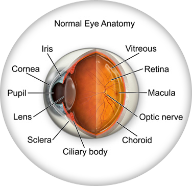
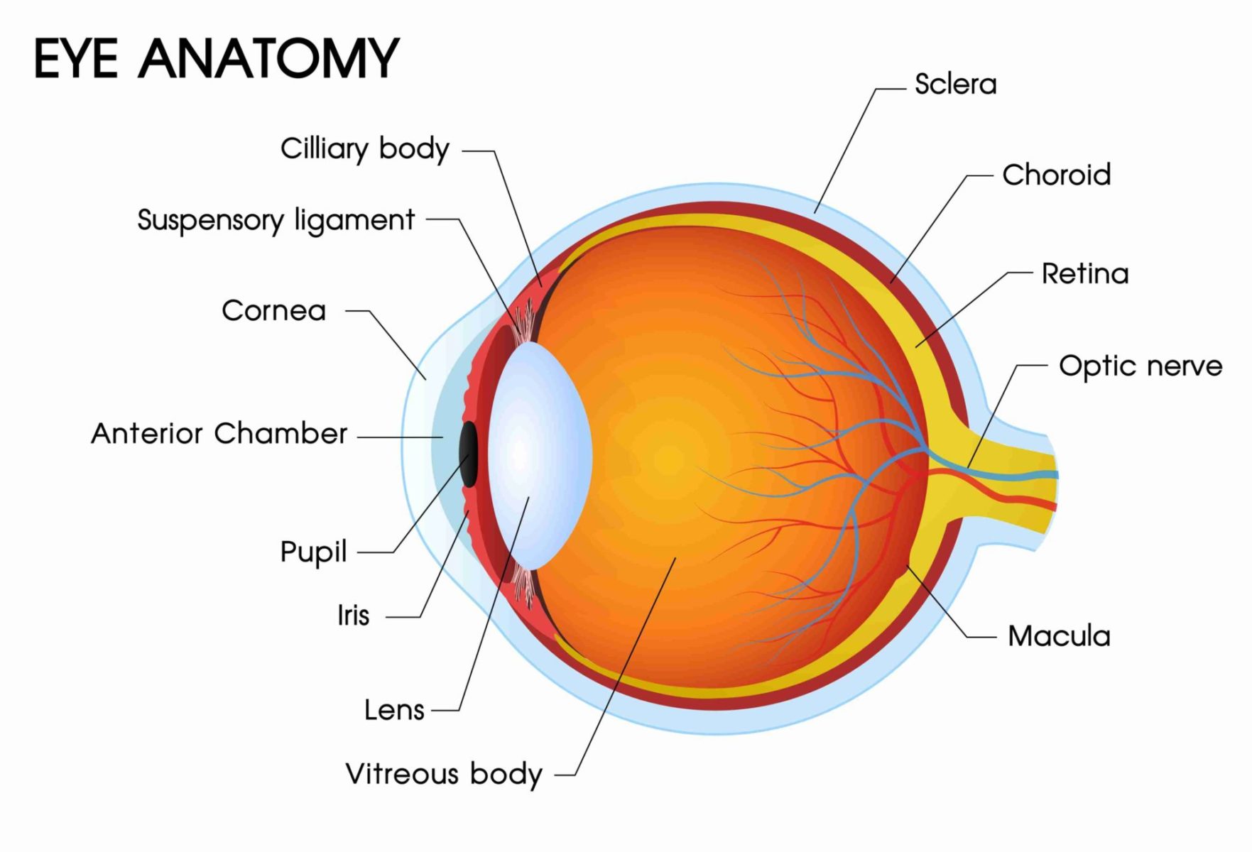
Eye Anatomy: Parts of the Eye and How We See - American ... Mar 9, 2021 · Eye Anatomy: Parts of the Eye Outside the Eyeball. The eye sits in a protective bony socket called the orbit. Six extraocular muscles in the orbit are attached to the eye. These muscles move the eye up and down, side to side, and rotate the eye. The extraocular muscles are attached to the white part of the eye called the sclera. This is a strong layer of tissue that covers nearly the entire surface of the eyeball. The Eyes (Human Anatomy): Diagram, Optic Nerve, Iris ... - WebMD Iris: the colored part Cornea: a clear dome over the iris Pupil: the black circular opening in the iris that lets light in Sclera: the white of your eye Conjunctiva: a thin layer of tissue that covers the entire front of your eye, except for the cornea
Eye anatomy: Muscles, arteries, nerves and lacrimal gland ... Oct 3, 2022 · Eye anatomy Bones of the orbit. The bony orbit is made out of seven bones, which include the maxilla, zygomatic bone, frontal bone,... Eyelid anatomy. The eyelids are soft tissue structures that cover and protect the anterior surface of the eyeball. ... Lacrimal gland. The lacrimal gland is a part ...
Eye anatomy
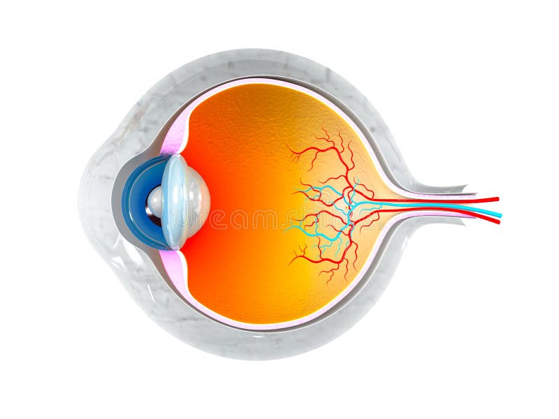
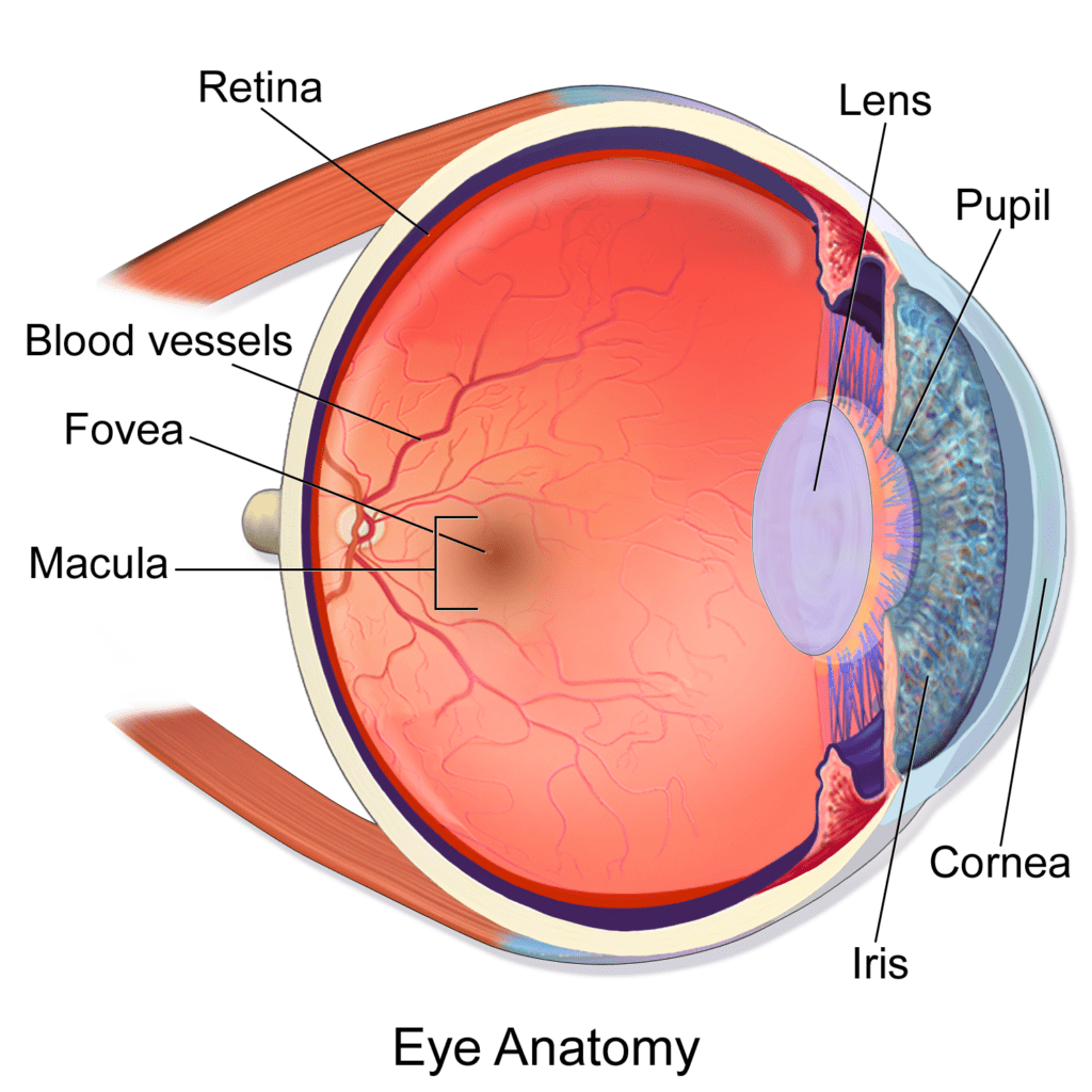
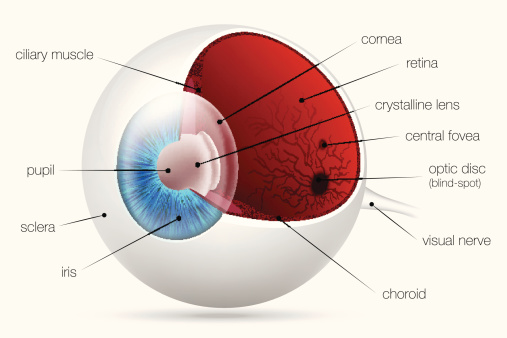
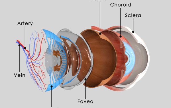
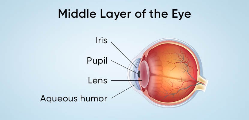
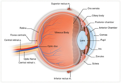
Eye Anatomy: Parts of the Human Eye - Vision Center Feb 3, 2023 · It is located underneath the white part of the eye (the sclera) and is composed of three parts: The iris Ciliary body Choroid Anatomy of the Eye | Kellogg Eye Center | Michigan Medicine Anatomy of the Eye Choroid Layer containing blood vessels that lines the back of the eye and is located between the retina (the inner... Ciliary Body Structure containing muscle and is located behind the iris, which focuses the lens. Cornea The clear front window of the eye which transmits and ... Anatomy of the Eye | Johns Hopkins Medicine Anatomy of the Eye Anterior chamber. . The front section of the eye's interior where aqueous humor flows in and out, providing nourishment... Aqueous humor. . The clear watery fluid in the front of the eyeball. Blood vessels. . Tubes (arteries and veins) that carry blood to and from the eye. ...
Eye anatomy. Eye Anatomy: A Closer Look At the Parts of the Eye Feb 27, 2019 · In a number of ways, the human eye works much like a digital camera: Light is focused primarily by the cornea — the clear front surface of the eye, which acts like a camera lens. The iris of the eye functions like the diaphragm of a camera, controlling the amount of light reaching the back of the... ... Anatomy of the Eye | Johns Hopkins Medicine Anatomy of the Eye Anterior chamber. . The front section of the eye's interior where aqueous humor flows in and out, providing nourishment... Aqueous humor. . The clear watery fluid in the front of the eyeball. Blood vessels. . Tubes (arteries and veins) that carry blood to and from the eye. ... Anatomy of the Eye | Kellogg Eye Center | Michigan Medicine Anatomy of the Eye Choroid Layer containing blood vessels that lines the back of the eye and is located between the retina (the inner... Ciliary Body Structure containing muscle and is located behind the iris, which focuses the lens. Cornea The clear front window of the eye which transmits and ... Eye Anatomy: Parts of the Human Eye - Vision Center Feb 3, 2023 · It is located underneath the white part of the eye (the sclera) and is composed of three parts: The iris Ciliary body Choroid
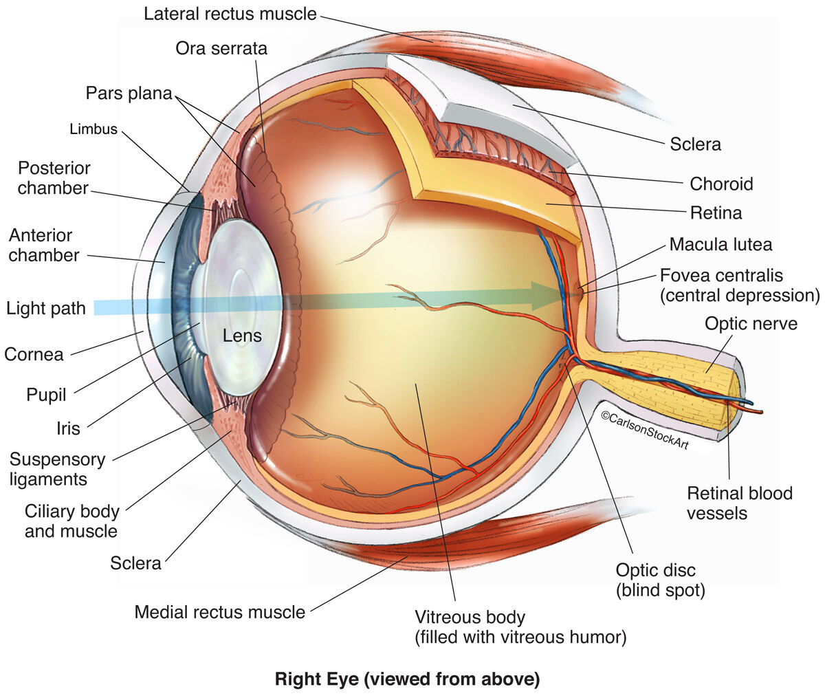
Eye Anatomy 1 Illustration | Carlson Stock Art
Anatomy of the Eye | Kellogg Eye Center | Michigan Medicine
Eye Anatomy explaining pupil and cornea

Anatomy of the Eye - Diagnosis 101
Vision and the eye's anatomy | HealthEngine Blog

File:Human eye anatomy.jpg - Wikimedia Commons
External anatomy of the human eye (with labels Stock Photo ...
3,131 Eye Anatomy Stock Photos - Free & Royalty-Free Stock ...

Retina of the Eye (Anatomy, Functions & Associated Conditions)
Eye - Anatomy, Functions, Diseases, Diagnosis, Tips For Good ...

Eye Anatomy - Vision Options by The Vision Council - Find an ...
Anatomy of the Human Eye | Dr. John Goosey

Eye Anatomy and How the Eye Works - Davidson Eye Associates
Eye Anatomy - Understand how your eyes work to produce one of ...
![Figure, Anatomy of the eye, showing...] - PDQ Cancer ...](https://www.ncbi.nlm.nih.gov/books/NBK65754/bin/CDR0000543553.jpg)
Figure, Anatomy of the eye, showing...] - PDQ Cancer ...
Anatomy of the Eye | MSVI

Anatomy of The Eye | Pappin & Pretorius Optometrists
Eye anatomy hi-res stock photography and images - Alamy
Anatomy of the Eye - StoryMD

Parts of the Eye - Eye Anatomy -Look After Your EyesLook ...
A Detailed Look at the Eye | The Canadian Association of ...
Eye Anatomy: Image Details - NCI Visuals Online
Eye Anatomy | Boroondara Eye Care

Eye Anatomy and How the Eye Works

Eye Anatomy - Bell Booth Sirkka Fabris Optometrists

About Your Eyes | Eye Anatomy | Greater Edmonton Eyecare
Eye Anatomy: Parts of the Eye and How We See - American ...
Eye Anatomy | Alberta Retina Consultants

ANATOMICAL DIAGRAM HUMAN EYE ANATOMY OPTOMETRIST CHART PRINT PREMIUM POSTER

About Basic Eye Anatomy | GEM Clinic - Glaucoma & Eye ...
Amazing Anatomy: Parts of the Eye Defined | Michigan Eye ...
Eye Anatomy - Exeter Eye

Eye Anatomy - Pediatric Ophthalmology PA

Vektor Stok Diagram Human Eye Anatomy Label Illustration ...
Your Eye Anatomy | Coast Optometry | Huntington Beach
Anatomy of the Eye – Columbia Eye Clinic

Human eye | Definition, Anatomy, Diagram, Function, & Facts ...
Eye Anatomy | Retina Specialists Orlando | Central Florida Retina

Eye Anatomy
Eye Anatomy: Parts of the Eye | Discount Contacts
Eye Anatomy - INTERNATIONAL SPECIALIST EYE CENTRE MALAYSIA
Eye Anatomy
Eye Anatomy: The 9 Main Parts of the Eye | Specialty Eye ...

The external anatomy of the eye - Stock Image - F002/4037 ...
Post a Comment for "44 eye anatomy"