39 microscope with label and function
The Best Firestick Remote Replacement 2022: Expert's Reviews - HomeChit Please Note: This is an infrared Remote. It DOES NOT have voice control function. This remote DOES NOT work with fire tv stick/fire tv box/firestick. New CT-RC1US-21 Infrared IR Replacement Remote Control Applicable for Toshiba Fire TV 43LF621U21 55LF621U21 TF-32A710U21 43LF421U21 50LF621U21 32LF221U21more Difference Between Mold and Yeast - Microbiology Info.com Hi, I am a medical physicist with experience in radiation protection. EM radiation does not cause yeast overgrowth. It does cause some heating if strong enough and so could contribute to creating a warm moist environment beneficial to yeast, but the strength from a phone mast 30m away would be way too low to do this.
Online Labs for schools - Developed by Amrita Vishwa Vidyapeetham and ... The development of OLabs includes the study and use of mathematical techniques to demonstrate the various complex functions in diverse areas of science. The labs make use of cutting edge simulation technology to create real world lab environments. Thorough study and research is done by research personnel for better understanding of the ...

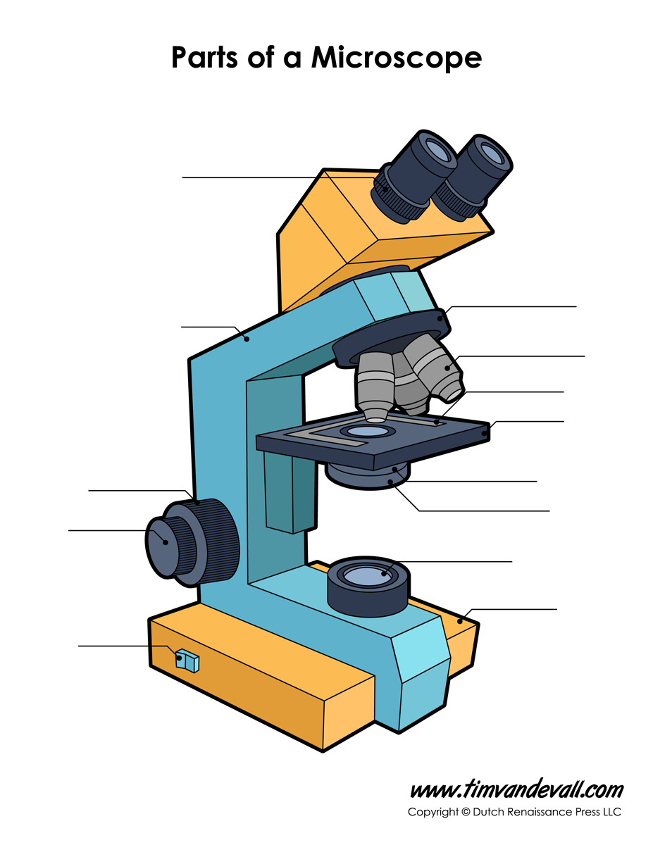
Microscope with label and function
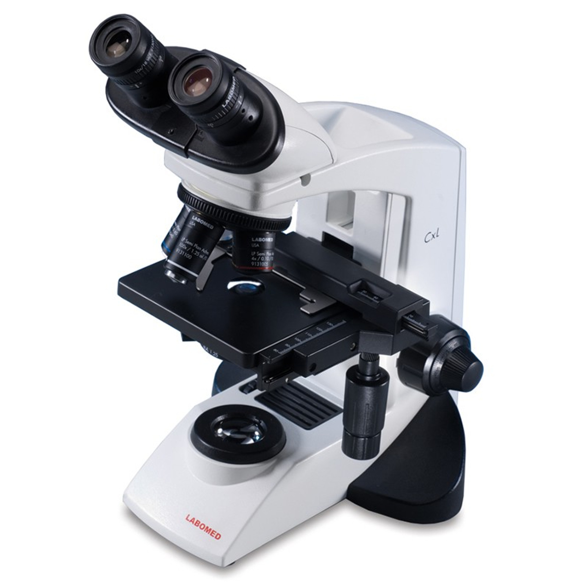
Microscope Diagram - cell division of e coli with continuous media flow ... Microscope Diagram - 15 images - give a well labelled diagram of compound microscope using of typical, bio tem biological transmission electron microscope university, labelled microscope diagram gcse micropedia, a compound microscope diagram micropedia, Medico hub Microscope MICROSCOPE : It is the most indispensable instrument in a biology laboratory. It helps to increase the res… Light Microscope (Theory) - Amrita Vishwa Vidyapeetham Microscope. Microscope is an optical instrument that uses lens or combination of lens to produce magnified images that are too small to seen by unaided eye. Microscope provides the enlarged view that helps in examining and analyzing the image. Microscope can be separated into optical theory microscopes (Light microscope), electron microscopes (eg.TEM, SEM) and scanning probe microscopes. (eg.AFM, PSTM).
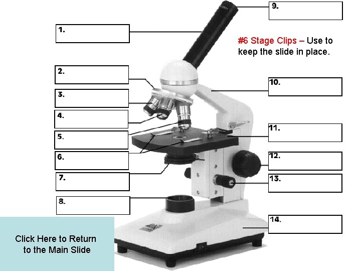
Microscope with label and function. Light Microscope (Procedure) - Amrita Vishwa Vidyapeetham A microscope slide is placed into the stage; clip it onto the mechanical stage. The nosepiece is rotated to the lowest-power objective i.e., 4x objective lens (objective lens with red band). The iris diaphragm is adjusted to the largest diameter, allowing the greatest amount of light to pass through. A Role for Cell 'Antennae' in Managing Dopamine Signals in the Brain Microscope image of a cultured mouse neuron from the striatum region of the brain labeled with a green fluorescent antibody that detects dopamine receptor 1. The receptor localizes along the cell surface and is enriched in a primary cilium projecting from the cell body. Nuclei are indicated in blue. Credit: of Kirk Mykytyn Uncovering the secret of insulin growth factor ternary complex The cryogenic transmission electron microscope (cryo-EM), data hub, and supercomputing resources of IBS were used to obtain its high-resolution three-dimensional molecular structure. It was... Fluorescence In Situ Hybridization (FISH) - Genome.gov The fluorescently labeled DNA finds its matching segment on one of the chromosomes, where it sticks. By looking at the chromosomes under a microscope, a researcher can find the region where the DNA is bound because of the fluorescent dye attached to it. This information thus reveals the location of that piece of DNA in the starting genome.
Function And Of Quiz Microscope Parts - tyg.bluservice.terni.it Parts of the Microscope and Their Function A microscope is a laboratory instrument used to examine objects that are too small to be seen by the naked eye STUDY the parts of the scope on the lab handout below Directions: Answer with well-written sentences In microscope: Mechanical components In microscope: Mechanical components. . Parasite reliance on its host gut microbiota for nutrition and survival ... D BODIPY and DAPI labeled images of fat body cells from 3LL GN D. melanogaster larvae inoculated with A. pomorum only, A. pomorum and Bacillus. sp., or A. pomorum, Bacillus. sp. and Rhodococcus sp ... Hypodermis of the Skin Anatomy and Physiology - Verywell Health Function The functions of the hypodermis include: 4 Fat and energy storage: Fat cells (adipocytes) make up the fatty (adipose) tissue that stores energy for the body. The hypodermis also helps creates hormones such as estrogen and leptin. 5 3 A role for cell 'antennae' in managing dopamine signals in the brain by Emily Caldwell, The Ohio State University Microscope image of a cultured mouse neuron from the striatum region of the brain labeled with a green fluorescent antibody that detects dopamine...
Understanding the wiring and programming of a labeler application The first input to highlight is the high-speed processor-in-the-loop (PIL) latch input updating at 5 microseconds (ms) or less for the label. The second input is the D1_01 (Digital Input 01), which updates at 600 ms for the product. Depending on the application and voltage, users should select the proper input. Worksheet Parts And Microscope Quizlet Use in addition to labeling the microscope parts, students are asked to describe the function of each piece of the optical microscope in his study of cells, the nancy larson science 4 curriculum actually teaches students the parts of the microscope and how to use it biology questions and answers discussion of the brain, and how it works, can be a … Microarray Technology - Genome.gov Definition 00:00 … Microarray technology is a general laboratory approach that involves binding an array of thousands to millions of known nucleic acid fragments to a solid surface, referred to as a "chip." The chip is then bathed with DNA or RNA isolated from a study sample (such as cells or tissue). DNA sequencing - Wikipedia DNA sequencing is the process of determining the nucleic acid sequence - the order of nucleotides in DNA.It includes any method or technology that is used to determine the order of the four bases: adenine, guanine, cytosine, and thymine.The advent of rapid DNA sequencing methods has greatly accelerated biological and medical research and discovery.
13 parts of the Compound Light Microscope Diagram | Quizlet
The Iris: Anatomy, Function, and Treatment - Verywell Health The iris sits in the uveal tract, which is the eye's middle layer. The iris lies in front of the lens and behind the cornea. It is made up of the following parts: 13. Iris pigment epithelium contains melanin granules and chromatophores that make up the eye color. Dilator and sphincter muscles that expand and contract to control the amount of ...

Light Microscopy: Function and Utility
ECLIPSE Ti2 Series | Inverted Microscopes - Nikon Europe B.V. The ECLIPSE Ti2 inverted microscope delivers an unparalleled 25mm field of view (FOV) that revolutionizes the way you see. With this incredible FOV, the Ti2 maximizes the sensor area of large-format CMOS cameras without making compromises, and significantly improves data throughput. The Ti2's exceptionally stable, drift-free platform is designed to meet the demands of super-resolution imaging while its unique hardware-triggering capabilities enhance even the most challenging, high-speed ...

Microscope and Function
Light-driven single-cell rotational adhesion frequency assay To overcome the limits of existing assays for cell adhesion measurements, we develop a light-driven single-cell rotational adhesion frequency assay (scRAFA), which enables label-free and sub-cellular-resolution quantification of adhesion of almost any targeted individual cells in clinical solutions (Fig. 1 d).

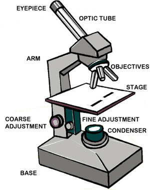
Compound Microscope Drawing With Parts and Functions
Multiparametric quantitative phase imaging for real-time, single cell ... For example, incubator-housed microscope systems for measuring real-time cell proliferation (e.g., Incucyte) have been shown to yield results concordant to CellTiter-Glo and BH3 profiling 18,19,20 ...

A diagram showing all of the parts of a compound light ...
Probiotics from Dairy Products on Intestinal Barrier Function Using ... After being treated with 10 8 CFU/ml Lactobacillus bulgaricus, Lactobacillus acidophilus, and Streptococcus thermophilus for 24 hours, the β-tubulin of Caco-2 cells was labeled by immunofluorescence method, and the green light of β-tubulin was observed under laser confocal microscope. Immunofluorescence method is a method that combines immunological methods (antigen-antibody specific binding) with fluorescent labeling technology to study the distribution of specific protein antigens in cells.

1 Body tube 2 Monocular Microscope Parts Functions
Multifunctional Laser Imaging of Cancer Cell Secretion with Hybrid ... In article number 2100734, Yu-Cheng Chen and colleagues have developed a label-free laser emission microscope for imaging secreted molecules associated with the cell-cell environment. Liquid crystal microdroplets are designed as signal amplifiers to report subtle molecular functions in a hybrid Fabry-Pérot microcavity.

PARTS OF MICROSCOPE| LEARN TO LABEL COMPOUND MICROSCOPE| JUST ...
N-STORM | Super-Resolution Microscopes - Nikon Instruments Inc. The 3D-Stack function allows multiple 3D STORM images from different Z positions to be captured and stitched into one image to create thicker STORM images. Tubulin of BSC-1 cell labeled with Alexa Fluor ® 647 Tenfold improvement of lateral resolution up to 20nm

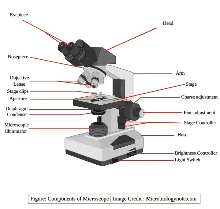
Parts of a microscope with functions and labeled diagram
3D Laser Scanning Microscope, VK-X200 - KEYENCE Combines the capabilities of an optical microscope, scanning electron microscope and roughness gauge into a single system. Provides non-contact profile, roughness and thickness measurements on nearly any material. Download the catalog for more information: VK-X Series 3D Laser Scanning Confocal Microscope Catalog [PDF:5.88MB] All (132) Support (40)

Parts of Microscope, Function, Names & Labeled Diagram ...
N.C. DPH: State Lab > Laboratory Improvement Training Workshops - NCDHHS In this basic level course, participants learn the principles of the brightfield microscope and the practical use of the precision instrument. Participants will develop skills in microscopy covering the following areas: Identify all parts of a microscope and understand the function of each. Clean and maintain the microscope.
Microscope Diagram Labeled, Unlabeled and Blank | Parts of a ...
Calibration Forms: Top 3 [Free Download] - SafetyCulture Calibration records include critical information such as reference design, tolerance limits, and calibration date and results. This information is used by technical services to ensure worker's compliance and assign immediate actions to mitigate quality risks.Regular equipment calibration helps the organization prevent accidents and damage that can lead to legal action and profit loss.

Label the microscope — Science Learning Hub
Gram Staining: Principle, Procedure, Interpretation, Examples and Animation Gram Staining is the common, important, and most used differential staining techniques in microbiology, which was introduced by Danish Bacteriologist Hans Christian Gram in 1884. This test differentiate the bacteria into Gram Positive and Gram Negative Bacteria, which helps in the classification and differentiations of microorganisms.

Parts of a microscope with functions and labeled diagram
Light Microscope (Theory) - Amrita Vishwa Vidyapeetham Microscope. Microscope is an optical instrument that uses lens or combination of lens to produce magnified images that are too small to seen by unaided eye. Microscope provides the enlarged view that helps in examining and analyzing the image. Microscope can be separated into optical theory microscopes (Light microscope), electron microscopes (eg.TEM, SEM) and scanning probe microscopes. (eg.AFM, PSTM).
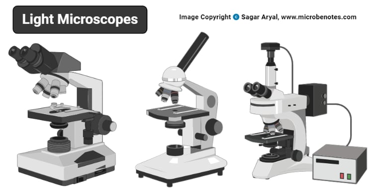
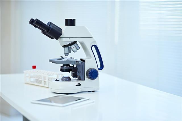
Light Microscope- Definition, Principle, Types, Parts ...
Medico hub Microscope MICROSCOPE : It is the most indispensable instrument in a biology laboratory. It helps to increase the res…
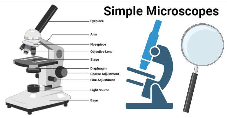
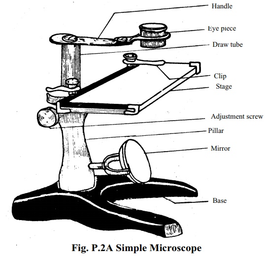
Simple Microscope- Definition, Principle, Magnification ...
Microscope Diagram - cell division of e coli with continuous media flow ... Microscope Diagram - 15 images - give a well labelled diagram of compound microscope using of typical, bio tem biological transmission electron microscope university, labelled microscope diagram gcse micropedia, a compound microscope diagram micropedia,
Comparing and Contrasting the Different Parts of the Microscope

Microscope With Labels Clip Art at Clker.com - vector clip ...

List: Parts of a Microscope and their Function | Pathwooded

Simple Microscope Definition, Magnification, Parts And Uses

Compound Microscope Parts and Functions Diagram | Quizlet
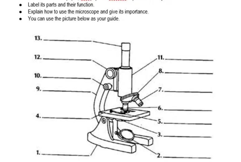
Answered: Label its parts and their function.… | bartleby

Compound Microscope Parts, Functions, and Labeled Diagram ...
Parts of a Microscope and Their Functions

Compound Microscope Parts, Functions, and Labeled Diagram ...
Parts of a Microscope and Their Functions

The Compound Microscope parts & how they work

what is the function of the pointer on a microscope

Compound Microscope Parts, Function, & Diagram | What is a ...
Free Microscope Drawing, Download Free Microscope Drawing png ...

Compound Microscope: Parts of Compound Microscope

Lasec Education | Key parts of a compound microscope and how ...

Label the microscope — Science Learning Hub

Compound Microscope- Definition, Labeled Diagram, Principle ...

microscope | Types, Parts, History, Diagram, & Facts | Britannica
Compound Microscope Parts, Functions, and Labeled Diagram ...

Compound Microscope Parts – Labeled Diagram and their ...
Parts of the Microscope and Their Function On
Microscope: Structure, Uses, Functioning Processes of Simple ...
Understanding the Compound Microscope Parts and its Functions ...
Parts of a microscope with functions and labeled diagram
Post a Comment for "39 microscope with label and function"