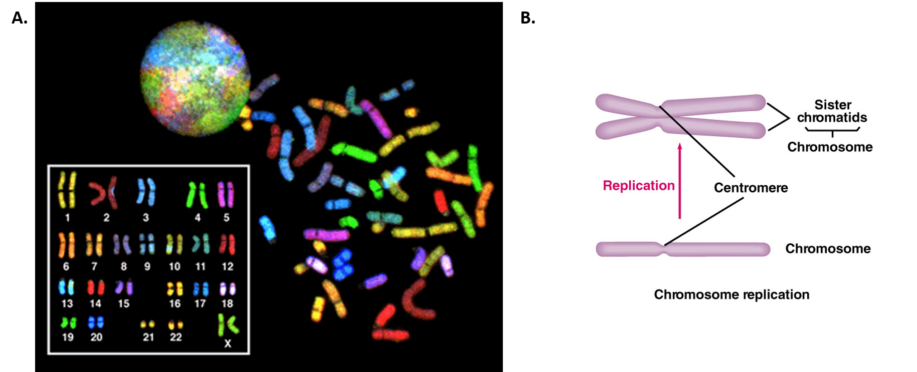
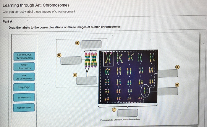
45 can you correctly label these images of chromosomes
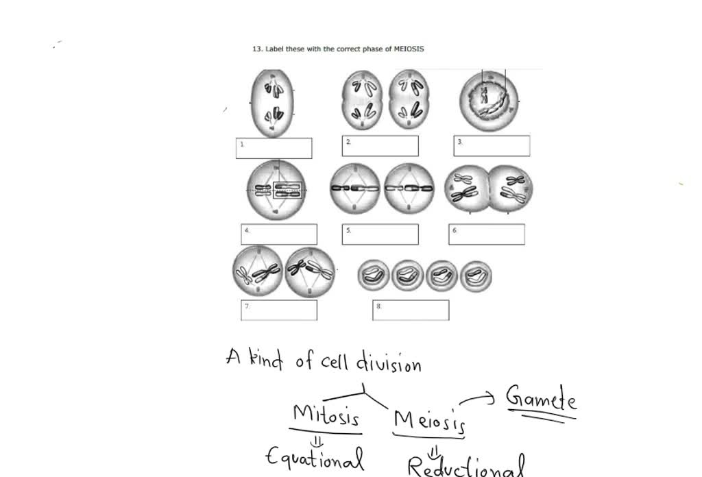
Answered: 2. Label the cell division photos. 1.… | bartleby Label the cell division photos. 1. Identify the stage of mitosis. 3. Identify the stage of mitosis. 4. Identify the dark- stained structures. 2. Identify small lines. PDF Meiosis picture labeling - WPMU DEV Chromosomes line up along equator, not in homologous pairs 21. Crossing-over occurs 22. Chromatids separate 23. Homologs line up alone equator 24. Cytoplasm divides, 2 daughter cells are formed 11. A cell with a diploid number of 20 undergoes meiosis. This will produce _____ daughter cells, each with _____ chromosomes. 12.
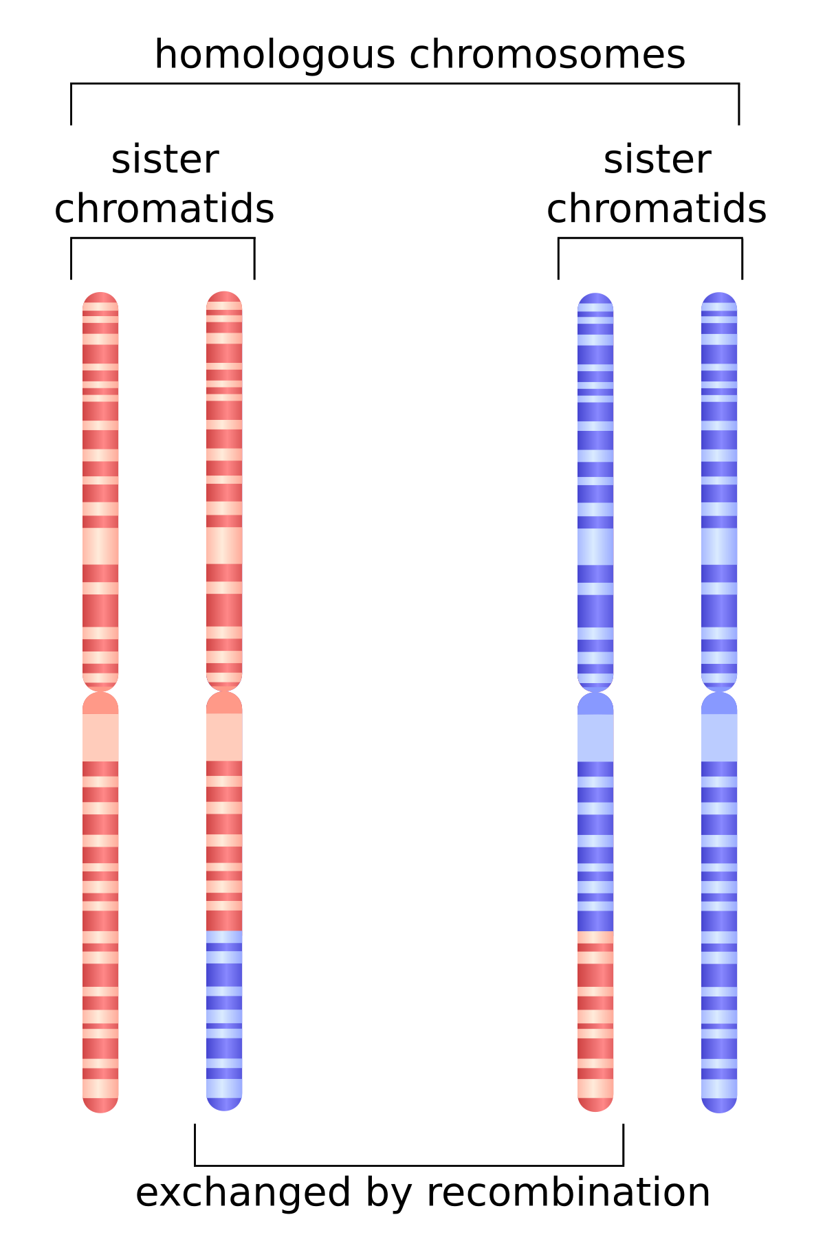
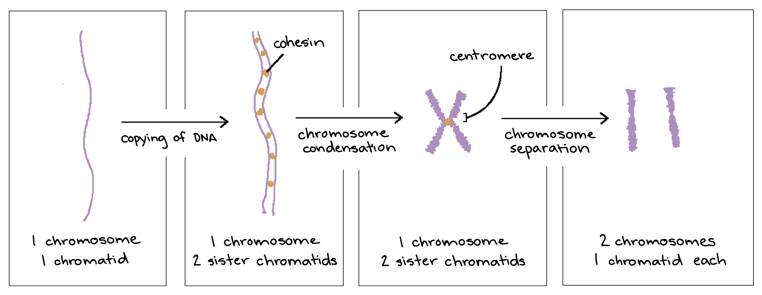
Chapter 8 Question 33 Multiple Choice Part A Two chromosomes in a ... ANSWER: Learning through Art: Chromosomes Can you correctly label these images of chromosomes? Part A Drag the labels to the correct locations on these images of human chromosomes. ANSWER:sister chromatids. homologous chromosomes. complementary chromosomes. 1 2 22 23

Can you correctly label these images of chromosomes
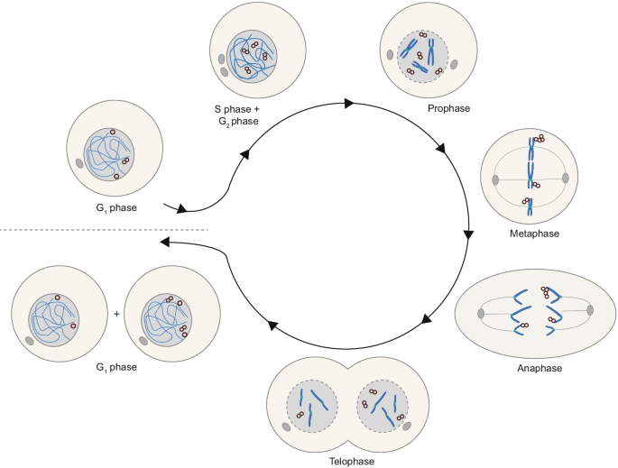
ch 8 mastering biology Flashcards - Quizlet Drag the labels onto the diagram to identify the stages of the cell cycle. 1. most of the cells life is spent in interphase 2. in phosphase microtubules form the mitototic spindle 3. at metaphase, the mitotic spindle is fully formed 4. in anaphase, sister chromatides separate 5. in telophase chromosomes become less condenced DOC Mitosis: Labeled Diagram - West Branch High School Four stages can be described for each nuclear division. First division of meiosis . Prophase 1: Each chromosome dupicates and remains closely associated. These are called sister chromatids. Crossing-over can occur during the latter part of this stage. Metaphase 1: Homologous chromosomes align at the equatorial plate. Anaphase 1 PDF Cell Division Animal Cell and Mitosis Key - Scarsdale Public Schools 1. Label the four phases of mitosis in the diagram. 2. Label the spindles and centrioles in one of the phases. 3. Color each chromosome in prophase a different color. Follow each of these chromosomes through mitosis. Show this by coloring the correct structures in each phase of mitosis. les Interphase Cytokinesis hase Chromatin Chromosome /(/t ...
Can you correctly label these images of chromosomes. A Labeled Diagram of the Animal Cell and its Organelles Chromosomes that determine the sex of an individual are known as sex chromosomes. In humans, X and Y are sex chromosomes. Females have two X chromosomes and males have one X and one Y chromosome. Autosomes are all the other chromosomes in the organism. Of the 46 chromosomes in humans, 44 are autosomes and the remaining two are the sex chromosomes. Classifying Chromosome Images Using Ensemble ... - SpringerLink Medical imaging processing has become an important diagnostic and therapy assistant tool. Chromosomes carry the genetic information of the human and the healthy human cell contains 23 pairs of chromosomes, 22 pairs of autosomes, and one pair of sex chromosomes (XX for female and XY for male) [].Changes in chromosomes number or structure do occur on rare occasions and maybe a sign of a genetic ... Answer correct chapter 8 question 15 multiple choice 6/27/22, 7:13 PM Chapter 8 31/48 Correct This is the order of the main four stages of mitosis. Pro means first. Meta means together. One of the definitions for ana is "divided into equal quantities." Telo means last. Learning through Art: Chromosomes Can you correctly label these images of chromosomes? 6 Main Parts of a Chromosome - Biology Discussion The following points highlight the six main parts of a chromosome. The parts are: 1. Pellicle and Matrix 2. Chromatids, Chromonema and Chromomeres 3. Centromeres 4. Secondary Constriction 5. Satellite 6. Telomere. Part # 1. Pellicle and Matrix: A membrane which surrounds each chromosome is said as pellicle.
Solved Can you label these chromosomes with the correct - Chegg Expert Answer. Who are the experts? Experts are tested by Chegg as specialists in their subject area. We review their content and use your feedback to keep the quality high. 100% (47 ratings) Transcribed image text: Can you label these chromosomes with the correct genetic terms? Drag the terms to their correct locations on the figure below. Chromosomes (article) | Cell cycle | Khan Academy The 46 chromosomes of a human cell are organized into 23 pairs, and the two members of each pair are said to be homologues of one another (with the slight exception of the X and Y chromosomes; see below). Human sperm and eggs, which have only one homologous chromosome from each pair, are said to be haploid ( 1n ). Chromosome Abnormalities Fact Sheet - Genome.gov In both processes, the correct number of chromosomes is supposed to end up in the resulting cells. However, errors in cell division can result in cells with too few or too many copies of a chromosome. Errors can also occur when the chromosomes are being duplicated. Other factors that can increase the risk of chromosome abnormalities are: Chromosome Mapping aka Ancestor Mapping - DNAeXplained Step 1 - Identify a common ancestor with those individuals you match on common DNA segments. This is really two steps, the common ancestor part, and the common DNA segment part. If these people are on your match list, we already know you have a common DNA segment over the vendor's match threshold.
Chapter 8 Homework Test Questions - StudyHippo.com question. Looking through a light microscope at a dividing cell, you see two separate groups of chromosomes on opposite ends of the cell. New nuclear envelopes are taking shape around each group. The chromosomes then begin to disappear as they unwind. You are witnessing. Click card to see the answer. answer. telophase. Types & Examples | Pros & Cons of Mutations - Bio Explorer Structural Chromosomal Mutations. This kind of chromosomal mutation usually occurs during any errors in cell division. This happens when homologous chromosomes paired up, genes in chromosomes broke apart, genes inserted in the wrong chromosome, or genes or set of genes are completely lost in the chromosome.. Basically, structural chromosomal mutations are classified into four: deletion ... Cell Division | Biology Quiz - Quizizz 30 seconds. Report an issue. Q. The diagram shows cells in different phases of mitosis. A student is looking for evidence that spindle fibers are separating the chromosomes to ensure that each new nucleus has one copy of each chromosome. Which cell is in the phase of mitosis that the student is searching for? answer choices. Cell 1. Cell 2. Answered: A-Mitosis 1. Draw and label the… | bartleby Label the chromatin as follows: long green- 1a short green- 2c long red- 1b short red- 2d 2- Let the cell pass S phase. Replicate or duplicate each chromatin fiber. Do this by getting another set of wire identical to the original set. Label as before. 3- Combine the replicated chromatin fiber using masking tape.
Mastering Biology 5 Flashcards - Quizlet Except during _____, cell division in humans results in daughter cells that have the same number of chromosomes and are genetically identical to each other and to the parent cell. ... Can you correctly label these images of chromosomes? After fertilization, the resulting zygote undergoes _____. mitosis.
PDF Name of Phase - WPMU DEV How many chromosomes would a diploid gorilla contain? 48 8. Are egg and sperm haploid or diploid? ... DIPLOID Part 2: Match the phase of meiosis with its correct description using the word bank provided. 1. 6. A Name of Phase Description ... On each of the images, label the phase of meiosis. 1. ANAPHASE II 2. CYTOKINESIS I 3. METAPHASE I
Chromosomes Fact Sheet - Genome.gov It is also crucial that reproductive cells, such as eggs and sperm, contain the right number of chromosomes and that those chromosomes have the correct structure. If not, the resulting offspring may fail to develop properly. For example, people with Down syndrome have three copies of chromosome 21, instead of the two copies found in other people.
(Get Answer) - Can you label these chromosomes with the correct genetic ... Learning through Art: Chromosomes Can you corectly label these images of chromosomes? Part A Drag the labels to the correct locations on these images of human chromosomes homologous chromosomes sister chromatids karyotype autosommes centromere?
Chromosome mutations | Biology Quiz - Quizizz Which of these is the correct order for the chromosome mutations in the image (from top to bottom)? Chromosome mutations DRAFT. 9th - 10th grade. 5 times. Biology. ... Non-disjunction involving the X chromosomes occurs during oogenesis and produces two kinds of eggs, XX and O (no X chromosomes). If normal sperms fertilize the two types of eggs ...
Which diagram most accurately shows the arrangement of homologous ... Each chromosome carries two clearly visible sister chromatids. Images in options C and D shows only single chromosome which can be the product of meiosis I, but the position of the second chromosome is not shown, which makes these images unclear. So, the correct answer is option A. Was this answer helpful? 0 0
Solved Learning through Art: Chromosomes Can you corectly - Chegg Question: Learning through Art: Chromosomes Can you corectly label these images of chromosomes? Part A Drag the labels to the correct locations on these images of human chromosomes homologous chromosomes sister chromatids karyotype autosommes centromere This problem has been solved! See the answer Show transcribed image text Expert Answer
Chapter 8 Homework - Free Essay Examples Database The function (s) of meiosis is/are _____. reproduction (production of gametes) Looking through a light microscope at a cell undergoing meiosis, you see that the chromosomes have joined into XX-shaped tetrads. These tetrads are lined up along a plane that runs through the center of the cell. This cell is in _____. meiosis I.
PDF Cell Division Animal Cell and Mitosis Key - Scarsdale Public Schools 1. Label the four phases of mitosis in the diagram. 2. Label the spindles and centrioles in one of the phases. 3. Color each chromosome in prophase a different color. Follow each of these chromosomes through mitosis. Show this by coloring the correct structures in each phase of mitosis. les Interphase Cytokinesis hase Chromatin Chromosome /(/t ...
DOC Mitosis: Labeled Diagram - West Branch High School Four stages can be described for each nuclear division. First division of meiosis . Prophase 1: Each chromosome dupicates and remains closely associated. These are called sister chromatids. Crossing-over can occur during the latter part of this stage. Metaphase 1: Homologous chromosomes align at the equatorial plate. Anaphase 1
ch 8 mastering biology Flashcards - Quizlet Drag the labels onto the diagram to identify the stages of the cell cycle. 1. most of the cells life is spent in interphase 2. in phosphase microtubules form the mitototic spindle 3. at metaphase, the mitotic spindle is fully formed 4. in anaphase, sister chromatides separate 5. in telophase chromosomes become less condenced


















Post a Comment for "45 can you correctly label these images of chromosomes"