40 drag each label into the appropriate position to identify what cell type is described by the label
Answered: Drag the labels to the appropriate… | bartleby Transcribed Image Text: Part A Drag the labels to the appropriate location in the figure. Reset Help Simple squamous epithelium Simple columnar epithelium Simple cuboidal epithelium Pseudostratified ciliated columnar epithelium Transitional epithelium Stratified squamous epithelium O Typę here to search PDF Purdue University Department of Chemistry each characteristic's number in the appropriate blank. Unless otherwise stated, all of the amino acids are at physiological pH 7.4. There are more than one characteristic for many of the amino acids and 24 total answers each worth one point. There will be a deduction for incorrect answers, so don't fill in the lines with unnecessary ...
(Solved) - Drag each label into the proper position to identify the ... Drag each label into the proper ...
Drag each label into the appropriate position to identify what cell type is described by the label
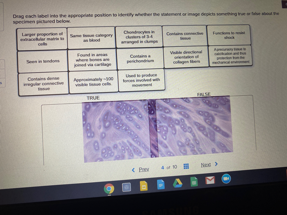
Answered: Drag each label into the appropriate… | bartleby Transcribed Image Text. Drag each label into the appropriate position to identify whether the statement or image depicts something true or false about the specimen pictured below. Larger proportion of extracellular matrix to Same tissue category as blood Chondrocytes in clusters of 3-4 Contains connective Functions to resist shock tissue cells ... Four Types of Protein Structure - ThoughtCo 2. Secondary Structure . Secondary Structure refers to the coiling or folding of a polypeptide chain that gives the protein its 3-D shape.There are two types of secondary structures observed in proteins. One type is the alpha (α) helix structure.This structure resembles a coiled spring and is secured by hydrogen bonding in the polypeptide chain. Anatomy and Physiology 2: Chapter 22: Immune System - Quizlet identify the type of hypersensitivity that occurs within 1-3 hours of exposure subacute hypersensitivities correctly order the events of inflammation 1. Release of chemicals 2. Vasodilation 3. Recruitment of immune cells 4. Delivery of plasma proteins redness, heat, and swelling are cardinal signs of _______ inflammation
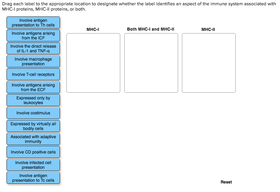
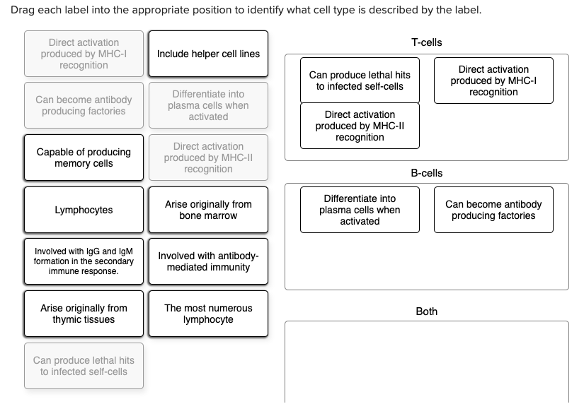
Drag each label into the appropriate position to identify what cell type is described by the label. Chapter 22 Homework Flashcards - Quizlet Drag each label into the appropriate position to identify what cell type is described by the label. 1. T-lymphocytes - mature in thymus - can produce lethal hits to infected self-cells - the most numerous lymphocyte - direct activation produced by MHC-II recognition - direct activation produced by MHC-I recognition 2. B-lymphocytes Label the heart - Science Learning Hub Drag and drop the text labels onto the boxes next to the diagram. Selecting or hovering over a box will highlight each area in the diagram. In this interactive, you can label parts of the human heart. Drag and drop the text labels onto the boxes next to the diagram. Selecting or hovering over a box will highlight each area in the diagram. Labeled Neuron Diagram | Science Trends Neurons are a type of cell and are the fundamental constituents of the nervous system and brain. Neurons take in stimuli and convert them to electrical and chemical signals that are sent to our brain. There are 3 major kinds of neurons in the spinal cord: sensory, motor, and interneurons. Neurons communicate vie electrical signals produced by ... [Expert Answer] Instructions: Drag each label to the ... - Brainly.com English High School answered • expert verified Instructions: Drag each label to the correct location on the image. Identify each passage as either functional text or expository text. Link to assignment photo below Expert-verified answer NFucini A functional text is written to fulfil a particular need or solve a problem.
PDF BIO 113 LAB 1. Anatomical Terminology, Positions, Planes, and Sections ... divide it up into smaller areas for study. The medical scheme divides the abdominal surface (and the abdominopelvic cavity deep to it) into four approximately equal regions called quadrants, named according to their relative position-they are, right upper quadrant, right lower quadrant, left upper quadrant, and left lower quadrant (Figure 6). Solved Drag each label into the appropriate position to | Chegg.com See the answer Drag each label into the appropriate position to identify what cell type is described by the label. Show transcribed image text Expert Answer 80% (5 ratings) T- cells - Arise originally from thymic tissues - Direct activation produced by MHC-I recognition and MHC-II recognit … View the full answer redirect.prod.mastering.pearsoncmg.com All of these question types require you to position items into an area of the answer box. Answer these kinds of questions on a computer, not on a smartphone. Basic keyboard instructions (applies to most keyboards) Labeling questions Ranking questions Sorting questions Sentence completion (vocabulary) questions Labeling, ranking, sorting, or sentence completion questions All of these question types require you to position items into an area of the answer box. Answer these kinds of questions on a computer, not on a smartphone. Basic keyboard instructions (applies to most keyboards) Labeling questions. Ranking questions. Sorting questions. Sentence completion (vocabulary) questions.
Chapter 17 digestive Flashcards - Easy Notecards Drag each label into the appropriate position in order to identify which type of macromolecule is the target of each digestive enzyme or compound. 20. ... Identify the digestive organ shown in each image below. Then, for each label, decide which organ is being described. Drag and drop each description onto the appropriate image. 66. How to find, highlight and label a data point in Excel scatter plot Here's how: Click on the highlighted data point to select it. Click the Chart Elements button. Select the Data Labels box and choose where to position the label. By default, Excel shows one numeric value for the label, y value in our case. To display both x and y values, right-click the label, click Format Data Labels…, select the X Value and ... Cell Cycle - Definition And Phases of Cell Cycle - BYJUS A typical eukaryotic cell cycle is divided into two main phases:-Interphase. Also known as the resting phase of the cell cycle; interphase is the time during which the cell prepares for division by undergoing both cell growth and DNA replication. It occupies around 95% time of the overall cycle. The interphase is divided into three phases:- Diagram of Human Heart and Blood Circulation in It - New Health Advisor A heart diagram labeled will provide plenty of information about the structure of your heart, including the wall of your heart. The wall of the heart has three different layers, such as the Myocardium, the Epicardium, and the Endocardium. Here's more about these three layers. Epicardium
Grade 6 Science Practice Test Answer Key
PDF In this chapter, you will learn that - Pearson Within each skeletal muscle, the muscle fibers are grouped into fascicles (fas ′ĭ-klz; "bundles") that resemble bundles of sticks. Surrounding each fascicle is a layer of dense irregular connective tissue called perimy-sium (per″ĭ-mis′e-um; "around the muscle"). Endomysium. The endomysium (en″do-mis′e-um; "within

Cytometry Part A Template - Wiley
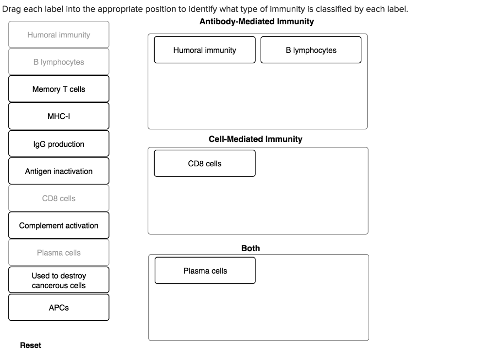
Drag each label into the appropriate position to identify what type of ... Drag each label into the appropriate position to identify what type of immunity is classified by each label. faith983 is waiting for your help. Add your answer and earn points. Expert-verified answer okpalawalter8 The categorization that shows the type of immunity is classified by each label. Under the Аntibоdy-Mediаted Immunity section:

Connect Homework Ch. 5 Flashcards | Quizlet
Label the Photomicrograph Using the Hints Provided. Drag each label into the appropriate position to identify what cell type is described by the label. Label the image of a compound light microscope using the terms provided. Label the testis and spermatic cord using the hints provided. Place the following pictures of white blood cells stained purple in the slides into the appropriate category.
Solved Drag each label into the appropriate position to ...
Human Karyotyping SE - bio - Name ... - StuDocu Create : Drag the chromosome to the appropriate position on the KARYOTYPING pane. Then select another chromosome, identify it, and place it on the karyotype. When you have identified and placed all of the chromosomes, click the camera ( ) to take a snapshot of the karyotype. Paste the snapshot into a document, and label it "Subject A ...

AHCDWeek2SOL10.pdf - 10. Award: 10.00 points Problems? Adjust ...
Solved Drag each label into the appropriate position to | Chegg.com Expert Answer Answer. The following are the labels that describe the T cell and B cells. 1. T-cell -Include helper cell lines. - Direct activation produced by MHC-1 recognition. -Direct activation produced by MHC-2 recognition - arise originally from t … View the full answer
Introduction to Chemistry
Drag each label into the appropriate position to identify what cell... Drag each label into the appropriate position to identify what cell type is described by the label. T cells Capable of developing Differentiate into memory cells plasma cells when activated Mature in thymus Lymphocytes Provide a defense B cell... Show more ... Show more Biology Science Anatomy Answer & Explanation Solved by verified expert

Label-free microfluidics for single-cell analysis - ScienceDirect
Parts of a microscope with functions and labeled diagram Ans. Microscopes are instruments that are used in science laboratories, to visualize very minute objects such as cells, and microorganisms, giving a contrasting image, that is magnified. Q. State functions of a microscope. Ans. A microscope is usually used for the study of microscopic algae, fungi, and biological specimens.

Post-Test - 3 Flashcards | Quizlet
Drag and drop into text question type - MoodleDocs Give each a label and drag the label. Scoring and Feedback All gaps are weighted identically. Only gaps that are filled correctly gain marks. There is no negative marking of gaps that are filled incorrectly. In interactive with multiple tries mode Combined feedback is shown after every try as well as when the question completes.

Chemical Analysis of Single Cells and Organelles | Analytical ...
Drag Each Label to the Location of Each Structure Described Drag each label to the location of each structure described. Place each label on the appropriate cerebral lobe. Drag and drop the text labels onto the boxes next to the heart diagram. Correctly label the following major systemic arteries. Each label can be used more than once.

Bio 2010 Exam 2 Flashcards | Quizlet
Anatomy and Physiology 2: Chapter 22: Immune System - Quizlet identify the type of hypersensitivity that occurs within 1-3 hours of exposure subacute hypersensitivities correctly order the events of inflammation 1. Release of chemicals 2. Vasodilation 3. Recruitment of immune cells 4. Delivery of plasma proteins redness, heat, and swelling are cardinal signs of _______ inflammation

Unit #1 Practice Questions (60 questions) Flashcards | Quizlet
Four Types of Protein Structure - ThoughtCo 2. Secondary Structure . Secondary Structure refers to the coiling or folding of a polypeptide chain that gives the protein its 3-D shape.There are two types of secondary structures observed in proteins. One type is the alpha (α) helix structure.This structure resembles a coiled spring and is secured by hydrogen bonding in the polypeptide chain.
PROSIDING
Answered: Drag each label into the appropriate… | bartleby Transcribed Image Text. Drag each label into the appropriate position to identify whether the statement or image depicts something true or false about the specimen pictured below. Larger proportion of extracellular matrix to Same tissue category as blood Chondrocytes in clusters of 3-4 Contains connective Functions to resist shock tissue cells ...

LOMBA FILM PENDEK - HIMAPIF 2022
Aerospace | Free Full-Text | Stereo Vision-Based Relative ...
Answered: Drag each label into the appropriate… | bartleby
Solved Drag each label to the appropriate location to | Chegg.com

File:2101 Blood Flow Through the Heart.jpg - Wikipedia

Connect Homework Ch. 5 Flashcards | Quizlet

AHCDW20Notes113.pdf - 113. Award: 1.00 point Problems? Adjust ...
Solved Drag each label into the appropriate position to ...
Informatics | Free Full-Text | Visual Analytics for ...
Drag and drop onto image question type - MoodleDocs

AHCDW20Notes113.pdf - 113. Award: 1.00 point Problems? Adjust ...

Chapter 22 Lymphatic System (Practice) Flashcards | Quizlet
Origin: Data Analysis and Graphing Software
Solved] Please see an attachment for details | Course Hero
Solved] Drag each label into the appropriate position to ...

Ch. 22 Assessment Flashcards | Quizlet

Connect Homework Ch. 5 Flashcards | Quizlet
Label-free imaging and classification of live P. falciparum ...

Electrocardiography - Wikipedia

AHCDW24Notes57.pdf - 57. Award: 1.00 point Problems? Adjust ...

Connect Homework Ch. 5 Flashcards | Quizlet

AHCDWeek2SOL10.pdf - 10. Award: 10.00 points Problems? Adjust ...

Drag each label to the correct location on the image ...

Brodart Just-A-Fold 12" archival plastic book jacket cover ...

AHCDW20Notes115.pdf - 115. Award: 1.00 point Problems? Adjust ...

Solved Drag each label into the appropriate position to ...
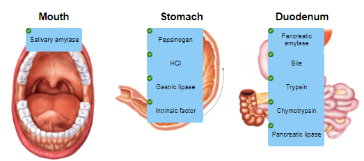
Chapter 17 digestive Flashcards - Easy Notecards

Connect Homework Ch. 5 Flashcards | Quizlet
Post a Comment for "40 drag each label into the appropriate position to identify what cell type is described by the label"