38 dental x-ray tube head diagram
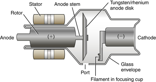
PDF Easy Guide to Dental X-ray Positioning - AAHA Ai m at the area between finger and thumb. Line up the bottom line on the tubehead to the canine tooth 103-101 & 203-201 (R or L incisors) 45 -50° Dependent upon the headtype of patient - tube head parallel to the nose crack 103-203 (All incisors) 45-60° Parallel to nose crack Mandible PDF The X-ray Tube - austincc.edu The diagram on the right shows the x-ray tube by itself . There are three major components that we will be discussing: The cathode which is negatively charged. Note its position on the diagram above. The Anode which is positively charged. And the Glass Envelope which supports the anode and cathode structures. 1
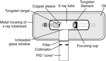
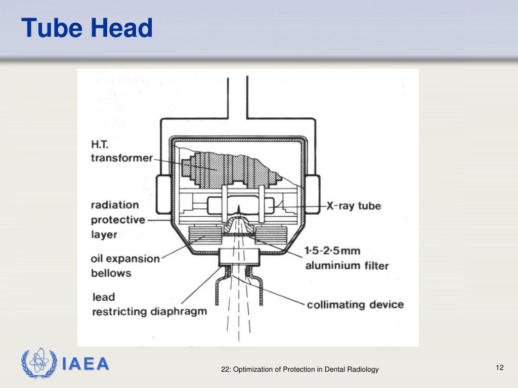
Dental X-ray Tube Head Diagram - local dentist Dental X-ray Tube Head Diagram. The dental x-ray technician should never receive primary radiation from a dental The following diagram will identify the location of these two devices (see figure 1-8). Tube head assembly: filter, collimator (diaphragm), PID or cone or tube.
Dental x-ray tube head diagram
Production Of X-Rays - Welcome to Dental Radiography Inside the metal tube housing is the x-ray tube. The diagram in figure 1-2 represents a dental x-ray tube head and a dental x-ray tube. This tube emits radiation in the form of photons (photons will be discussed in Lesson 2) or x-rays. X-ray photons expose the film. In addition to exposing the film, it also exposes the patient to radiation. Production of Dental X-Rays - Dental Radiology - YouTube A summary of how an xray is produced in the dental xray tube head.Information taken from:Dental Radiography Principle and Techniques - by Iannucci and Howert... Types of Dental X-Rays and Why You Need Them A dental x-ray is the common term for a dental radiograph. It is one of the dentist's most important diagnostic tools, giving him or her a better picture of what's going on with your teeth than simply looking in your mouth. Dental radiographs work by using a small, controlled burst of radiation to create a picture of the tooth.
Dental x-ray tube head diagram. PDF INSTALLATION INSTRUCTIONS - Belmont Dental Equipment Company x-ray tube, high voltage generator or both. B. Duty cycle A cool down interval of 50 seconds or more must be allowed between each 1 second exposure. (a 25 second cool down must be allowed between each 0.5 second exposure.) This will avoid the accumulation of excess heat and prolong the tube head life. C. Tube head cooling curve 0 0 PDF Intraoral radiographic techniques The Periapical radiograph (IOPA) is the basic investigation that gives graphic information about the alveolar bone, periodontal areas and the hard tissues of the tooth. Each image usually shows 2-4 teeth. Indications The clinical indications include: 1. To visualize Periapical region 2. detection of apical infection/inflammation. 3. Panoramic Dental X-ray - Radiologyinfo.org Panoramic radiography, also called panoramic x-ray, is a two-dimensional (2-D) dental x-ray examination that captures the entire mouth in a single image, including the teeth, upper and lower jaws, surrounding structures and tissues. The jaw is a curved structure similar to that of a horseshoe. US4157476A - Dental X-ray tube head - Google Patents In a dental x-ray tube head, the x-ray tube is in a casing that supports the tube and shields against stray radiation being projected to the environment through the housing of the tube head. The...
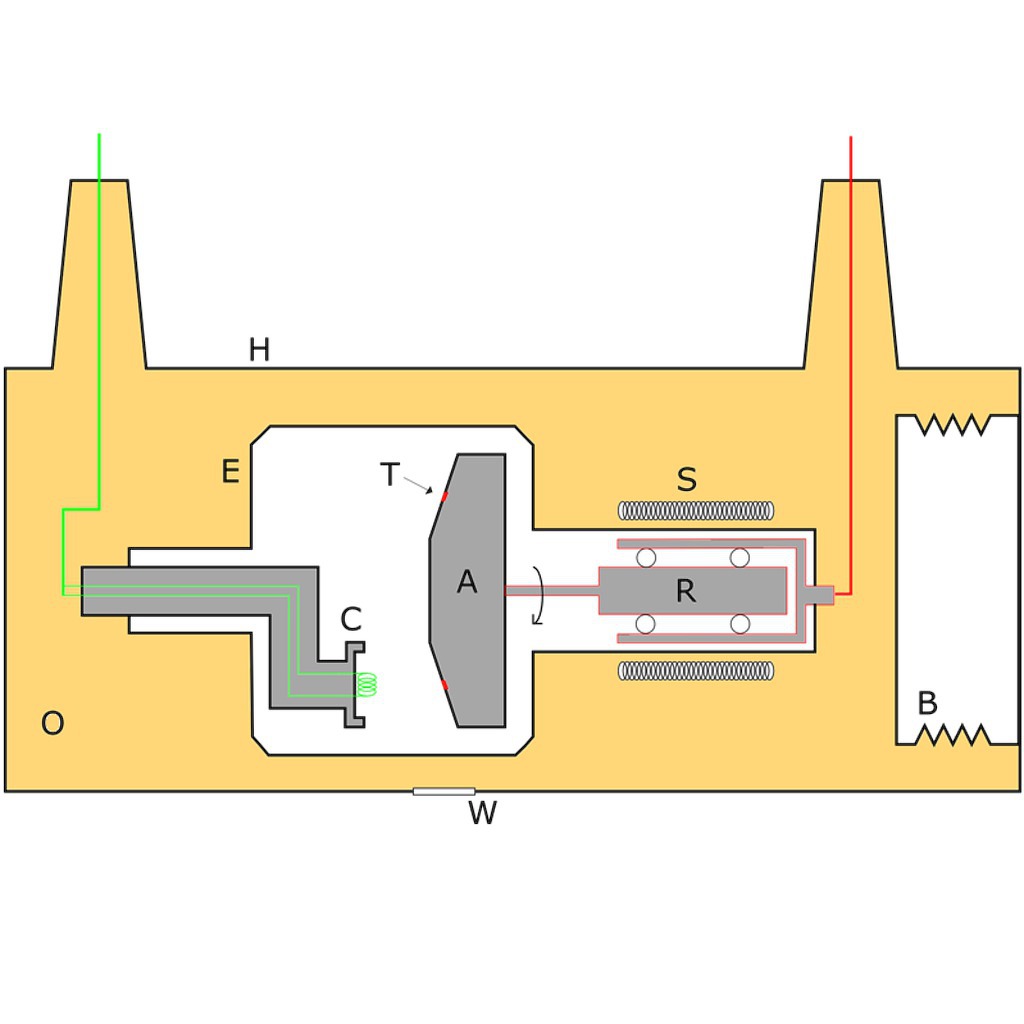
Steps In The Process Of Xray Production - Dental Radiography See figures 1-3, 1-4, and 1-5 for a diagram of the complete procedure. Figure 1-3. Tube head with the filament of the cathode emitting electrons. Figure 1-4. Electrons speeding toward the anode (tungsten target). Leaded Glass Tube Metal Housing Cathode Anode (Tungsten Target) Cathode Anode (Tungsten Target) Aluminum Filter Dental Radiology Practice Test Questions And Answers - ProProfs A-P view of the skull. 12. In the construction of an X-ray tube, the function of a step-down transformer is to. A. Convert the line current of 110 volts to less than 10 million amperes. B. Convert the line current of 110 volts to less than 10 volts. C. Convert the line current of 220 volts to less than 10 volts. PDF MODEL 097 DENTAL X-RAY - Belmont Equipment x-ray tube, high voltage generator or both. B. Duty cycle A cool down interval of 50 seconds or more must be allowed between each 1 second exposure. (a 25 second cool down must be allowed between each 0.5 second exposure.) This will avoid the accumulation of excess heat and prolong the tube head life. C. Tube head cooling curve 1. X-ray tube - Wikipedia X-ray tube. A modern dental x-ray tube. The heated cathode is on the left. Centre is the anode which is made from tungsten and embedded in the copper sleeve. An X-ray tube is a vacuum tube that converts electrical input power into X-rays. [1] The availability of this controllable source of X-rays created the field of radiography, the imaging of ...
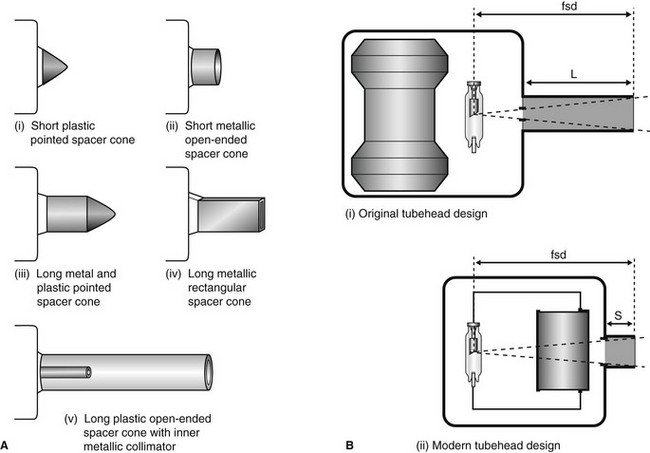
3: Dental X-ray equipment, image receptors and image ... - Pocket Dentistry B Diagrams showing the original tubehead design with the X-ray tube at the front of the head, thus requiring a long spacer cone (L) to achieve a near-parallel X-ray beam and the correct focus to skin distance (fsd) and the modern tubehead design with the X-ray tube at the back of the head, thus requiring only a short spacer cone (S) to achieve ... Labelling Dental X-Ray Tube head Diagram | Quizlet Start studying Labelling Dental X-Ray Tube head. Learn vocabulary, terms, and more with flashcards, games, and other study tools. Scheduled maintenance: Saturday, June 5 from 4PM to 5PM PDT Radiation Protection - Welcome to Dental Radiography The following diagram will identify the location of these two devices (see figure 1-8). Figure 1-8. Tube head assembly: filter, collimator (diaphragm), PID or cone or tube. ... short wavelength x-rays (photons) to pass through the filter. Filters on dental x-ray machines with over 70 kVp have a minimum thickness of 2.5 mm of aluminum. Those ... HDX Intra-oral X-ray - Intra-oral - X-ray - Equipment Flow Dental HDX Wall-Mount X-Ray Machine reduces patient x-ray exposure time by roughly 50 percent compared to conventional systems, while its high definition produces razor sharp images. ... Rated Peak Tube Potential: 65 kVDC: Rated Tube Current: 7 mA: Line Voltage Range: 100-130/200-250 50/60 Hz: Line Voltage Regulation: 4%: Rated Line ...
The X-ray Tube - Radiology Key The general-purpose x-ray tube is an electronic vacuum tube that consists of an anode, a cathode, and an induction motor all encased in a glass or metal enclosure (envelope). Figure 5-3 provides a labeled illustration of this design. Recall that the anode is the positive end of the tube and the cathode is the negative end of the tube.
schematic diagram x ray machine Block Diagram X Ray Machine - Wiring Diagram Schema wiring88.blogspot.com. xrf. Section Ii Production Of Xrays 16 Parts And Components Of The Dental . machine parts dental xray ray panel control tube head diagram xrays figure components section production ii representation extension arm radiography
LECTURE 1: The Tube Head - Intro Dental Radiography I LECTURE 1: The Tube Head The Dental X-ray Tube The filament is heated by the filament current. Electrons are emitted by the hot filament and travel to the anode. This flow of electrons from the...
Dental X-ray Tubehead Diagram - Quizlet piece of lead that reshapes the size of the beam and further filters out low-wavelength beams PID Position Indicator Device: device that is lined with lead and contains an aperture through which the primary x-ray beam passes as it leaves the device and heads to the patient Copper Stem (anode) positive electrode Focusing Cup (cathode)
PDF Dental X-ray 097 - ホーム the x-ray tube, high voltage generator or both. B. Duty cycle A cool down interval of 50 seconds or more must be allowed between each 1 second exposure. (a 25 second cool down must be allowed between each 0.5 second exposure.) This will avoid the accumulation of excess heat and prolong the tube head life. C. Tube head cooling curve 1.
How does a dental x ray tube head work? - Answers Inside the metal tube housing is the x-ray tube. The diagram in figure 1-2 represents a dental x-ray tube head and a dental x-ray tube. This tube emits radiation in the form of photons (photons...
Dental X-ray Tube Head How It Works - local dentist Dental X-ray Tube Head How It Works. The tubehead is a sealed, heavy metal housing that contains the x-ray tube that has a specific function that contributes to the safe exposure of dental x-rays. the safety features, will allow you to explain to the patient how x-rays work. Dental X-ray Tube Head How It Works. How does the procedure work?
PDF DENTAL X-RAY 097 - Belmontdental the x- ray tube, high voltage generator or both. B. Duty cycle A cool down interval of 50 seconds or more must be allowed between each 1 second exposure. (a 25 second cool down must be allowed between each 0.5 second exposure.) This will avoid the accumulation of excess heat and prolong the tube head life. C. Tube head cooling curve 1.
X ray Tube head, its components & functions - YouTube This video will help you in understanding the different parts in an x-ray tube head and its functions
Label the Dental X-ray Tubehead (Screencast) - Wisc-Online OER Label the Dental X-ray Tubehead (Screencast) By Joan Rohrer. The tubehead is a sealed, heavy metal housing that contains the x-ray tube that produces dental x-rays. This learning object will provide students with practice identifying and labeling the dental x-ray tubehead. Related.
Dental X-ray tube head - General Electric Company Dental x-ray apparatus which includes the new shielding construction is depicted in FIG. 1. The dental x-ray tube head is generally designated by the reference numeral 10. It comprises a housing 11 having a bottom wall 12 to which a tubular assembly 13 is attached. This assembly is otherwise known as a cone.
Types of Dental X-Rays and Why You Need Them A dental x-ray is the common term for a dental radiograph. It is one of the dentist's most important diagnostic tools, giving him or her a better picture of what's going on with your teeth than simply looking in your mouth. Dental radiographs work by using a small, controlled burst of radiation to create a picture of the tooth.
Production of Dental X-Rays - Dental Radiology - YouTube A summary of how an xray is produced in the dental xray tube head.Information taken from:Dental Radiography Principle and Techniques - by Iannucci and Howert...
Production Of X-Rays - Welcome to Dental Radiography Inside the metal tube housing is the x-ray tube. The diagram in figure 1-2 represents a dental x-ray tube head and a dental x-ray tube. This tube emits radiation in the form of photons (photons will be discussed in Lesson 2) or x-rays. X-ray photons expose the film. In addition to exposing the film, it also exposes the patient to radiation.







![PDF] Tube angulation effect on radiographic analysis of the ...](https://d3i71xaburhd42.cloudfront.net/2474d7c2e22cba363573626935ca08ecc19e7744/3-Figure2-1.png)




Post a Comment for "38 dental x-ray tube head diagram"