41 label the photomicrograph of thin skin
PDF Name the Condition - Dr. Scott Croes' Website Name the 4 layers of thin skin in both the cartoon and the photomicrograph. Name the 4 layers of thin skin in both the cartoon and the photomicrograph. •Name the Layers of skin and label the dermal papilla and dermis •Name the Layers of skin and label the dermal papilla Sebaceous Gland Label The Photomicrograph Of Thin Skin / Accessory ... Sebaceous Gland Label The Photomicrograph Of Thin Skin / Accessory Structures Of The Skin Anatomy And Physiology. This is the spellchex dictionary for online spell checking. Endocrine mucin producing sweat gland carcinomas always categorical a minimum of one.
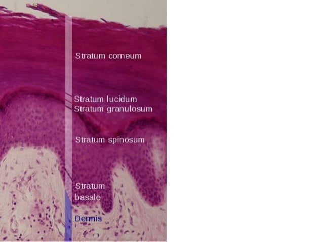
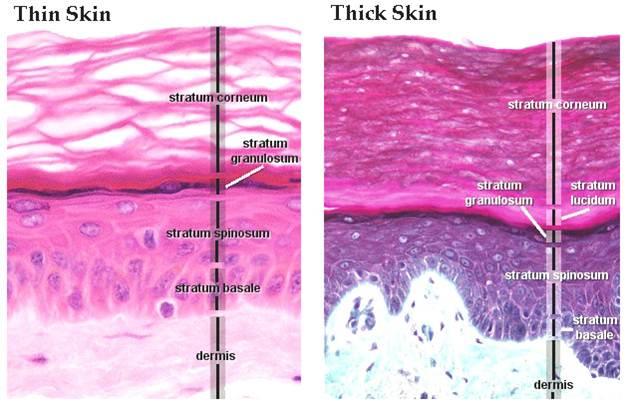
5.1 Layers of the Skin - Anatomy & Physiology "Thick skin" is found only on the palms of the hands and the soles of the feet. It has a fifth layer, called the stratum lucidum, located between the stratum corneum and the stratum granulosum ( Figure 5.1.2 ). Figure 5.1.2 - Thin Skin versus Thick Skin: These slides show cross-sections of the epidermis and dermis of (a) thin and (b) thick skin.
Label the photomicrograph of thin skin
skin labeling quizlet - gooddog.co.za Start studying photomicrographs of skin (thin skin). It is made up of three layers, the epidermis, dermis, and the hypodermis, all three of which vary significantly in their anatomy and function. This is why the scrotum is located outside of the body. Label The Photomicrograph Of Thin Skin. : Chapter 13, Page 3 ... Label The Photomicrograph Of Thin Skin. : Chapter 13, Page 3 - HistologyOLM. 28.09.2020 · this involves depositing a thin layer of heavy metal (eg, platinum) on the specimen by placing it in the path of a beam of metal ions in a vacuum. Label The Photomicrograph Of Thick Skin / Collagen Elastin Like ... Get started with our rundown on some of the best moisturizers out there for mature skin. Thick skin · stratum basale (also known as s. 1 answer to label the photomicrograph of thin skin. "thick skin" is found only on the palms of the hands and the soles of the feet. It has a fifth layer, called the stratum lucidum, located between the .
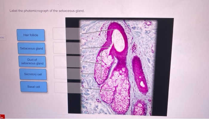
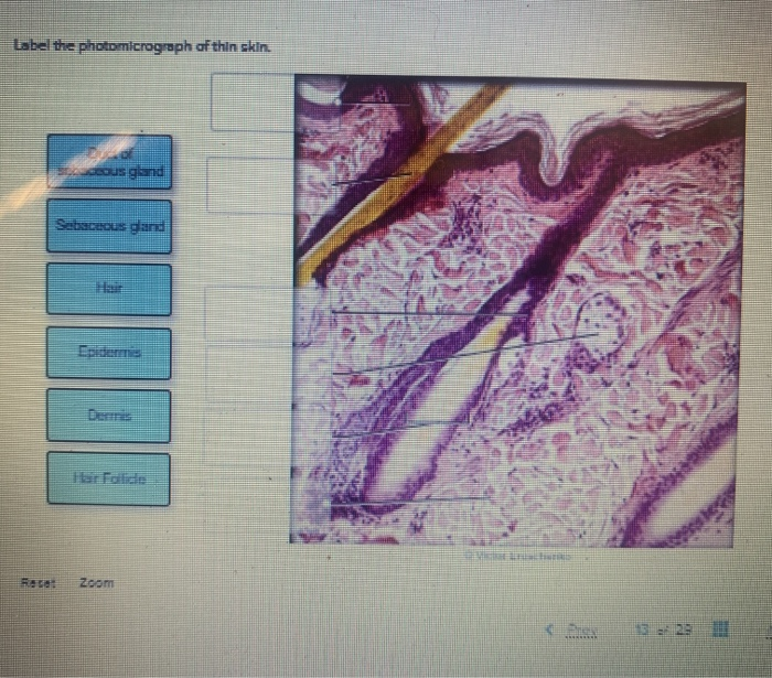
Label the photomicrograph of thin skin. Solved Label the photomicrograph of thin skin. Dermis Duct - Chegg Expert Answer. Who are the experts? Experts are tested by Chegg as specialists in their subject area. We review their content and use your feedback to keep the quality high. 100% (33 ratings) A …. View the full answer. Transcribed image text: Label the photomicrograph of thin skin. Dermis Duct of sebaceous gland Hair Follicle Sebaceous gland ... photomicrograph of thick skin Diagram - Quizlet Start studying photomicrograph of thick skin. Learn vocabulary, terms, and more with flashcards, games, and other study tools. Photomicrograph of Thin Skin Quiz - PurposeGames.com This is an online quiz called Photomicrograph of Thin Skin There is a printable worksheet available for download here so you can take the quiz with pen and paper. Your Skills & Rank Total Points 0 Get started! Today's Rank -- 0 Today 's Points One of us! Game Points 5 You need to get 100% to score the 5 points available Actions Anatomy, Skin (Integument), Epidermis - StatPearls - NCBI Bookshelf Skin is the largest organ in the body and covers the body's entire external surface. It is made up of three layers, the epidermis, dermis, and the hypodermis, all three of which vary significantly in their anatomy and function. The skin's structure is made up of an intricate network which serves as the body's initial barrier against pathogens, UV light, and chemicals, and mechanical injury ...
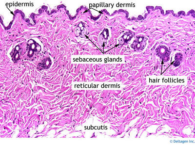
Label The Photomicrograph Of The Sebaceous Gland - Era Elang Label the photomicrograph of thin skin. If the gland become blocked, the sebum can be forced out into the dermis, where it elicits an inflammatory response. Label the photomicrograph of the skin and its accessory structures. Epidermis hair follicle duct of sebaceous gland sebaceous gland. How you will use this image and then you will be able to ... Junqueira's Basic Histology Text and Atlas, 14th Edition Enter the email address you signed up with and we'll email you a reset link. Full text of "NEW" - Internet Archive An icon used to represent a menu that can be toggled by interacting with this icon. unit 4 lab.docx - LAB Unit 4 EXERCISE 7: The ... - Course Hero FIGURE 7.2: Photomicrograph of the skin. epidermis (EPI-derm-is) • dermal papillae (puh-PILL-ee) • hypodermis (HY-poh- der-mis) • papillary (PAP-il-lary) layer of dermis • reticular layer of dermis 1. Dermal Papillae 2. Epidermis 3. Papillary layer of dermis 4. Reticular layer of dermis 5. Hypodermis
Label the photomicrograph in Figure 7.4. Examine a slide of hairy skin ... Label the layers of the skin on the diagram and the photograph. Be able to identify the layers on a microscope slide. Look at the skin slide under a microscope. a) Epidermis 1) Stratum corneum ii) Stratum lucidum 111) Stratum granulosum iv) Stratum... Posted 11 months ago Recent Questions in Basics of Statistics Q: PDF The Integumentary System - Holly H. Nash-Rule, PhD Label the skin structures and areas indicated in the accompanying diagram of thin skin. Then, complete the statements that follow. a. Lamellar granules contain glycolipids that prevent water loss from the skin. b. Fibers in the dermis are produced by fibroblasts . Effects of Disturbed Flow on Vascular Endothelium ... Jan 01, 2011 · Vascular endothelial cells (ECs) are exposed to hemodynamic forces, which modulate EC functions and vascular biology/pathobiology in health and disease. The flow patterns and hemodynamic forces are not uniform in the vascular system. In straight parts of the arterial tree, blood flow is generally laminar and wall shear stress is high and directed; in branches and curvatures, blood flow is ... (PDF) DiFiore's Atlas of Histology with Functional ... Enter the email address you signed up with and we'll email you a reset link.
35 Label The Photomicrograph Of The Sebaceous Gland. - Labels For You
A&P 1 Exercise_7 Activity 1 & 2 & RYK and UYK.docx - Course Hero Apocrine sweat Gland Label the photomicrograph in Figure 7.4. 1. Sebaceous glands 2. Hair follicle 3. Hair root 4. Hair bulb 5. Papilla of hair ... Translucent layer found in thick skin, absent in thin skin. Stratum Spinosum 6. Appears to have thorn-like projections in prepared slides. Reticular Region 7.
Solved: Label The Photomicrograph Of Thin Skin. Deous Glan... | Chegg.com
Physiology of the Endometrium and ... - Physiological Reviews Apr 29, 2020 · The physiological functions of the uterine endometrium (uterine lining) are preparation for implantation, maintenance of pregnancy if implantation occurs, and menstruation in the absence of pregnancy. The endometrium thus plays a pivotal role in reproduction and continuation of our species. Menstruation is a steroid-regulated event, and there are alternatives for a progesterone-primed ...
Biology Archive | January 15, 2017 | Chegg.com
Question : Question 31 points Label the photomicrograph of thin skin ... Question : Question 31 points Label the photomicrograph of thin skin. Hair Follicle : 391984. Question. 31 points Label the photomicrograph of thin skin. Hair Follicle Hair Dermis Sebaceous gland Duct of sebaceous gland Reset zoom. Solution. 5 (1 Ratings ) Solved. Biology 2 Years Ago 68 Views.

Thick Skin
Review on nanoparticles and nanostructured materials: history ... Apr 03, 2018 · Mollusk shells consists of “nacre”, which is a hierarchical nanocomposite. Nacre is designed by alternating micrometer-sized and sub-micrometer CaCO 3 aragonite platelets, which are separated by a thin layer of bio-macromolecular “glue”. Enhanced stiffness, impact resistance, strength, and toughness are some of the mechanical properties ...
Skin histology
label the layers of the epidermis in thick skin The epidermis of thin skin ranges from 0.07-0.15 . DermNet provides Google Translate, a free machine translation service. It is made of four or five layers of epithelial cells, depending on its location in the body. Start studying photomicrograph of thick skin. Skin checker . Hypodermis label the layers of the epidermis in thick skin in figure 7.2.
stratum-corneum - NYSCC
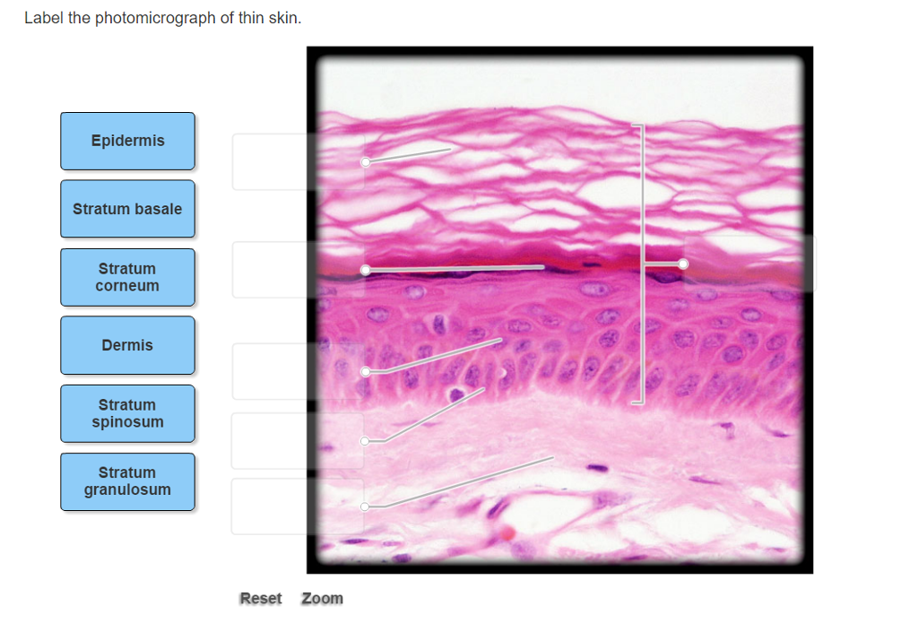
Layers of the Skin - Anatomy and Physiology Skin that has four layers of cells is referred to as "thin skin.". From deep to superficial, these layers are the stratum basale, stratum spinosum, stratum granulosum, and stratum corneum. Most of the skin can be classified as thin skin. "Thick skin" is found only on the palms of the hands and the soles of the feet.

Biology Archive | March 09, 2017 | Chegg.com
Anatomy and Physiology Homework Chapter 6 Flashcards - Quizlet Label the photomicrograph of thin skin.-Duct of sebaceous gland-Epidermis-Hair-Sebaceous gland-Dermis-Hair Follicle-Epidermis-Hair-Duct of sebaceous gland-Sebaceous gland-Hair Follicle-Dermis Explanation: Thin skin is located throughout the body. Refer to APR 3.0 for further information.

Thin Skin versus Thick Skin - Histology | Skin anatomy, Thick skin ...
Label The Photomicrograph Of Thin Skin Quizlet - Skin Labeling Review ... Label the photomicrograph of thin skin. Learn vocabulary, terms, and more with flashcards, games, and other study tools. C) contains more sweat glands than thin skin. In the diagram of skin shown below, which labeled structure generates fingerprints? Label the photomicrograph of thin skin. Start studying photomicrographs of skin labeling.
Integumentary System Histology - Skin (labels) - histology slide
(Solved) - Label the photomicrograph of thin skin. O Stratum granulosum ... Label the photomicrograph ...
Post a Comment for "41 label the photomicrograph of thin skin"