40 correctly label the structures associated with unmyelinated nerve fibers in the pns.
PDF Principles of Nerve Conduction Studies - AANEM the specific use described by the authors and are "off-label" (i.e. use not described on the product's label). ... motor NCS and NEE assess the motor nerve fibers of the PNS ... The motor and sensory axons composing the PNS may be myelinated or unmyelinated. The myelin does not coat the nerve fiber uniformly but, rather, in segments. ... Overview of neuron structure and function - Khan Academy Anatomy of a neuron. Neurons, like other cells, have a cell body (called the soma ). The nucleus of the neuron is found in the soma. Neurons need to produce a lot of proteins, and most neuronal proteins are synthesized in the soma as well. Various processes (appendages or protrusions) extend from the cell body.
Action potential - Definition, Steps, Phases | Kenhub An action potential is defined as a sudden, fast, transitory, and propagating change of the resting membrane potential. Only neurons and muscle cells are capable of generating an action potential; that property is called the excitability. This article will discuss the definition, steps and phases of the action potential.
Correctly label the structures associated with unmyelinated nerve fibers in the pns.
4. Neuroanatomy CNS.pdf - 4/25/2018 4. Neuroanatomy CNS 4 ... - Course Hero ANSWER: Correct The cerebral hemispheres, which form the superior part of the brain, account for about 83% of total brain mass.Nearly the entire surface of the cerebral hemispheres is marked by elevated ridges called sulci. The cerebral hemispheres account for about 83% of total brain mass. Parasympathetic Nervous System Functions - Simply Psychology Nerve fibres of the PSNS arise within the central nervous system. The primary nerves involved are cranial nerves. Below are some of the main cranial nerves in the PSNS: Vagus nerve - approximately 75% of all the parasympathetic nerves are vagus nerves. These nerves have branches in many key organs such as the stomach, kidneys, bladder, and ... The Peripheral Nervous System | SEER Training A connective tissue sheath called the epineurium surrounds each nerve. Each bundle of nerve fibers is called a fasciculus and is surrounded by a layer of connective tissue called the perineurium. Within the fasciculus, each individual nerve fiber, with its myelin and neurilemma, is surrounded by connective tissue called the endoneurium.
Correctly label the structures associated with unmyelinated nerve fibers in the pns.. Ch 14 Flashcards | Chegg.com Describe the main steps in the regeneration of a nerve fiber. Describe a typical chemical synapse in reference to its microanatomy and the role of the neurotransmitters. Define the following terms and correctly match with either the CNS or PNS: ganglia, nerve, center (or nucleus), tract (or fascicle), and column. Which of the following are effectors a receptors b A) Receptors B) Stimuli C) Reflexes D) Glands E) Sense organs. 19) What is another name for the autonomic nervous system? A) Visceral sensory division B) Somatic sensory division C) Visceral motor division D) Somatic motor division E) Central nervous system. 20) Nerves are __________ of the nervous system. A) organs. Myelin Sheath: What They Are, Their Function, & Damage From MS - WebMD The myelin sheath wraps around the fibers that are the long threadlike part of a nerve cell. The sheath protects these fibers, known as axons, a lot like the insulation around an electrical wire ... If a neuron is prevented from sending a neurotransmitter across a ... A)A Schwann cell folds its plasma membranearound several fibers. B)A Schwann cells wraps its plasma membranearound each individual fiber as it does with myelinated fibers. C)An oligodendrocyte cells wraps its plasmamembrane around each fiber as it does with myelinated fibers. D)Satellite cells cluster around each axon to form apseudo-myelin sheath.
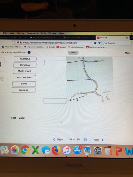
Lab 1 Homework BIOL 320 Flashcards | Quizlet Correctly label the structures associated with unmyelinated nerve fibers in the PNS. Indicate the specified region of the diencephalon. Thalamus Determine which general anatomic feature of the brain is illustrated in the figure. Sulcus Determine which specific tract is depicted in the figure. Muscle and Nervous Tissue Review Flashcards | Quizlet Label the structures of a skeletal muscle fiber. Label the structures of a sarcomere. Based on your understanding of the process of skeletal muscle contraction, indicate whether each figure represents a relaxed, partially contracted, or fully contracted skeletal muscle. Place the events involved in muscle contraction in the correct order. 1.) 16.1 Neurons and Glial Cells - Concepts of Biology - 1st Canadian Edition An axon is a tube-like structure that propagates the integrated signal to specialized endings called axon terminals. These terminals in turn synapse on other neurons, muscle, or target organs. Chemicals released at axon terminals allow signals to be communicated to these other cells. Chapter 12 QS Anatomy (Nervous System) Flashcards - Quizlet The structural classification of neurons is based on the number of processes that extend from the neuron cell body. Match these definitions to the correct term. 1. Many dendrites and a single axon (Click to select) 2. One dendrite and one axon (Click to select) 3.
An Integrated View on Neuronal Subsets in the Peripheral Nervous System ... In the gastrointestinal tract, intrinsic neurons are those whose cell bodies lie within the organ, whereas extrinsic nerves (e.g. sensory nerve fibers) have their cell bodies outside the innervated organ. Typically, the soma of extrinsic sensory afferents is located within dorsal root ganglia, celiac ganglia, superior or inferior mesenteric ... Unit 4 Anatomy & Physiology Flashcards - Quizlet The Schwann cell's plasma membrane spirals repeatedly around the unmyelinated fiber as it does in a myelin sheath. false place the following in the order that an electrical impulse would travel beginning with the post-synaptic membrane 1. dendrite 2. soma 3. axon hillock 4. internode 5. node of rangier 6. terminal aborization 7. synaptic knobs visceral anatomy example - dublinwestsda.ie Correctly label the structures associated with unmyelinated nerve fibers in the PNS. Tears are more common in spots that are vulnerable. The word usage examples above have been gathered from various sources to reflect current and historial usage. Peripheral Nervous System | histology The CNS consists of the brain and the spinal cord, while the PNS is composed of nerves and groups of nerve cells (neurons), called ganglia. The nerves of the PNS carry sensory (afferent) inputs to the CNS and motor (efferent) output from the CNS to the skeletal and cardiac muscles and the smooth muscles of blood vessels, organs and glands.
32 Correctly Label The Following Anatomical Features Of A Neuron ...
Anatomy Midterm Lecture Flashcards - Quizlet Correctly label the structures associated with unmyelinated nerve fibers in the PNS. ... and the ability to secrete a chemical that will stimulate the next cell when an electrical signal reaches the end of a nerve fiber. ... Correctly label the structures associated with the lacrimal apparatus.
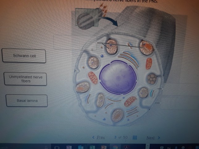
Solved: CORRECTLY LABEL THE STRUCTURES ASSOCIATED WITH UNM... | Chegg.com
23 some neurons are specialized to detect stimuli - Course Hero A Schwann cells wraps its plasma membrane around each individual fiber as it does with myelinatedfibers. An oligodendrocyte cells wraps its plasma membrane around each fiber as it does with myelinated fibers.Satellite cells cluster around each axon to form a pseudo-myelin sheath. A Schwann cell folds its plasma membrane around several fibers . 25.
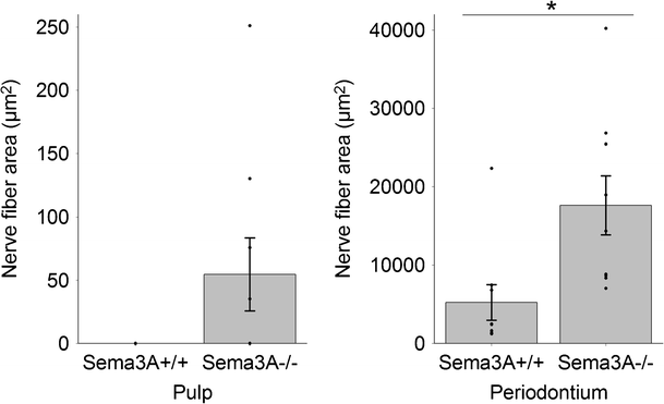
Sema3A chemorepellant regulates the timing and patterning of dental ...
lab 6 answers.docx - 201 LAB 6: NERVOUS SYSTEM With Answers 202 LAB 6 A ... 216 ear models - label using the following terms: oval window, cochlea, cochlear nerve, vestibular nerve, tympanic cavity, round window, vestibule, semicircular canal, tympanic membrane, stapes, malleus, incus, inner ear, middle ear, external ear, auricle or pinna, external auditory canal, tympanic membrane 8 new 9 auricle or pinna external …
Post a Comment for "40 correctly label the structures associated with unmyelinated nerve fibers in the pns."